Japanese
English
- 有料閲覧
- Abstract 文献概要
- 1ページ目 Look Inside
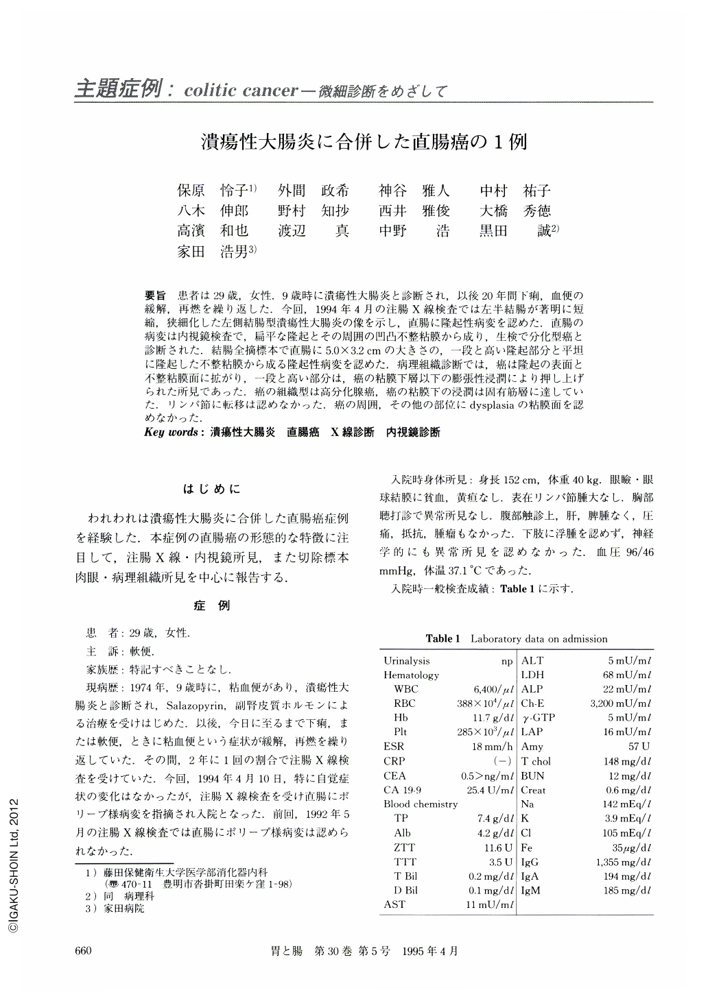
要旨 患者は29歳,女性.9歳時に潰瘍性大腸炎と診断され,以後20年間下痢,血便の緩解,再燃を繰り返した.今回,1994年4月の注腸X線検査では左半結腸が著明に短縮,狭細化した左側結腸型潰瘍性大腸炎の像を示し,直腸に隆起性病変を認めた.直腸の病変は内視鏡検査で,扁平な隆起とその周囲の凹凸不整粘膜から成り,生検で分化型癌と診断された.結腸全摘標本で直腸に5.0×3.2cmの大きさの,一段と高い隆起部分と平坦に隆起した不整粘膜から成る隆起性病変を認めた.病理組織診断では,癌は隆起の表面と不整粘膜面に拡がり,一段と高い部分は,癌の粘膜下層以下の膨張性浸潤により押し上げられた所見であった.癌の組織型は高分化腺癌,癌の粘膜下の浸潤は固有筋層に達していた.リンパ節に転移は認めなかった.癌の周囲,その他の部位にdysplasiaの粘膜面を認めなかった.
A 29-year-old woman had been suffering from relapsing, remitting symptoms with diarrhea and bloody stool for 20 years with the diagnosis of ulcerative colitis. Double contrast colonography in April, 1994 showed markedly shortened and narrowed left-side colon and polypoid lesion in the rectum. Endoscopy demonstrated flat protruded lesion with irregular uneven mucosa and biopsy specimen from this lesion revealed differentiated adenocarcinoma. The total-colectomy specimen showed a polypoid lesion consisting of protruded a lesion and a flat elevated area, measuring 5.0×3.2 cm in the rectum. The left-side colon and rectum were markedly shorted and narrowed with atrophic mucosa. Histological examination demonstrated cancerous mucosa spread on the surface of the protruded lesion and the surrounding irregular mucosa. In the protruded area, submucosal, expansive invasion of the cancer tissue had compressed the cancerous mucosa upward and formed a polypoid lesion. The histological type of cancer was well differentiated adenocarcinoma and the cancerous tissue had invaded as far as the proper muscle layer. No metastasis was seen in the expired lymph nodes. In the cancerous lesion and other examined colon and rectum mucosa, no dysplastic mucosa was found. The earlier lesion of this cancer was thought to be a flat cancer of the rectum.

Copyright © 1995, Igaku-Shoin Ltd. All rights reserved.


