Japanese
English
- 有料閲覧
PPV購入案内
1,320円 (1,200円+税10%) こちらは、19ページから30ページの文献です。購入には医書.jp本会員登録が必要です。
会員登録完了後に改めて上記「購入サイトへ」を選択してください。
または「購入サイトへ」選択後に「購入ログイン」、「新規会員登録」の順に進んでください。
会員登録について詳しくはをご確認ください。
- Abstract 文献概要
- 1ページ目 Look Inside
- 参考文献 Reference
- サイト内被引用 Cited by
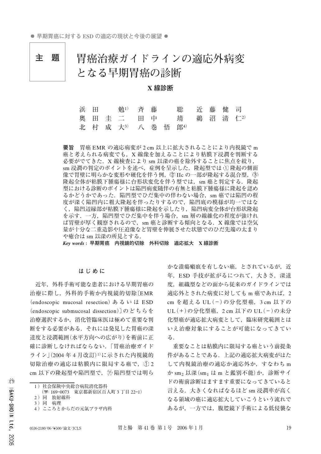
要旨 胃癌EMRの適応病変が2cm以上に拡大されることにより内視鏡でm癌と考えられる病変でも,X線像を加えることにより粘膜下浸潤を判断する必要がでてきた.X線検査によりsm以深の癌を除外することに焦点を絞り,sm浸潤の判定のポイントを述べ,症例を呈示した.隆起型では①隆起の側面像で胃壁に明らかな変形や硬化を伴う例,②IIcの一部が隆起する混合型,③隆起全体が粘膜下腫瘍様に台形状変化を伴う型では,sm癌と判定する.隆起型における診断のポイントは陥凹病変随伴の有無と粘膜下腫瘍様に隆起を認めるかどうかであった.陥凹型でひだ集中の伴わない場合,sm癌では陥凹の程度が深く陥凹内に粗大隆起を伴ったりするので,陥凹底の模様が均一ではなく,陥凹辺縁部が粘膜下腫瘍様に隆起を示したり,陥凹病変全体が台形状隆起を示す.一方,陥凹型でひだ集中を伴う場合,sm層の線維化の程度が強ければ胃壁が厚く観察されるので,sm癌と診断する傾向となる.X線像では空気量が十分な二重造影や圧迫像など胃壁を伸展させた状態でのひだ先端の太まりや癒合はsm以深の所見とする.
Endoscopic therapy has made it possible to remove early cancers within the guidelines such as cancers less than 2 cm in size, depressed type without ulceration and differentiated type. Recently, a new technique called ESD (endoscopic submucosal dissection) which can remove a wider range of cancers, has been introduced and it has been used to treat early cancers more than 2 cm in size, depressed type with ulceration and undifferentiated type. But, in such lesions which are considered as being beyond the indication of endoscopic therapy, cancerous infiltration is not always limited to the mucosa and sometimes has already invaded the submucosa. When we endoscopically remove a cancer it is hard to diagnose the depth of invasion relying only on endoscopy, so we emphasize that radiological figures are very useful for determining the depth of invasion.
The radiological findings which indicate cancerous invasion to the submucosa are as follows. In elevated lesions ; ( 1 ) rigidity of the gastric wall often pictured in the lateral view of the tumor, ( 2 ) complicated depressed change around the elevated lesion in the double contrast frontal view, ( 3 ) complicated margin just like submucosal tumor in the double contrast or compression view. In the depressed lesions ; ( 1 ) deep depression and complicated central nodural changes in the double contrast view, ( 2 ) elevated change covered with normal mucosa around the depressed lesion in the double contrast or compression view, ( 3 ) obvious clubbing and adhesion of converging folds in the double contrast view with an abundant amount of air.
Copyright © 2006, Igaku-Shoin Ltd. All rights reserved.