Japanese
English
- 有料閲覧
PPV購入案内
1,320円 (1,200円+税10%) こちらは、1617ページから1625ページの文献です。購入には医書.jp本会員登録が必要です。
会員登録完了後に改めて上記「購入サイトへ」を選択してください。
または「購入サイトへ」選択後に「購入ログイン」、「新規会員登録」の順に進んでください。
会員登録について詳しくはをご確認ください。
- Abstract 文献概要
- 1ページ目 Look Inside
- サイト内被引用 Cited by
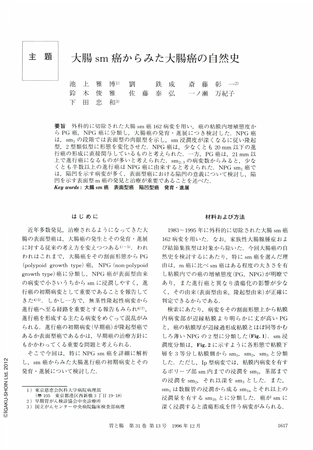
要旨 外科的に切除された大腸sm癌162病変を用い,癌の粘膜内増殖態度からPG癌,NPG癌に分類し,大腸癌の発育・進展につき検討した.NPG癌は,sm1の段階では表面型の肉眼型を示し,sm浸潤度が深くなるに従い隆起型,2型類似型に形態を変化させた.NPG癌は,少なくとも20mm以下の進行癌の形成に直接関与しているものと考えられた.一方,PG癌は,21mm以上で進行癌になるものが多いと考えられた.sm2,3の病変数からみると,少なくとも半数以上の進行癌はNPG癌に由来すると考えられた.NPGsm1癌では,陥凹を示す病変が多く,表面型癌における陥凹の意義について検討し,陥凹を示す表面型m癌の発見と治療が重要であることを述べた.
The development of colorectal cancer was studied by examining growth patterns in 162 lesions of resected submucosal invasive cancer. The lesions were classified into two groups ; polypoid growth (PG) and non polypoid growth (NPG), based on cross-section view. The degree of submucosal invasion was classified into three stages as sm1, sm2 and sm3.
Ninety four lesions (58.0%) were classified as PG, while 68 lesions (42.0%) as NPG. Mean size of NPG at the intramucosal part was smaller than that PG (13.2 mm and 22.7 mm, respetively) . All sm1 lesions of NPG had the appearance of superficial type cancer macroscopically and none of them were accompanied with ulcers. These findings indicated that NPG were derived from superficial type cancers.
Submucosal invasive NPG-type cancers tended to change their macroscopic features with the progress of submucosal invasion. Macroscopically, all sm1 lesion belonged to the superficial type, while sm2,3 lesions were superficial type, protruded type or small ulcerated type. Most NPG lesions seemed capable of developing into advanced carcinomas when their sizes were less than 20 mm in diameter, while PG lesions were considered able to develop into advanced cancers only when their sizes had become greater than 21 mm in diameter. The fact that most sm2,3 were associated with NPG lesions had us to suspect that more than 50% of advanced cancers arise from submucosal invasive NPG type lesions. The study of sm1 lesions showed that a depressive appearance of a lesion is important for indicating that submucosal invasion has begun.
Copyright © 1996, Igaku-Shoin Ltd. All rights reserved.