Japanese
English
- 有料閲覧
PPV購入案内
1,320円 (1,200円+税10%) こちらは、555ページから561ページの文献です。購入には医書.jp本会員登録が必要です。
会員登録完了後に改めて上記「購入サイトへ」を選択してください。
または「購入サイトへ」選択後に「購入ログイン」、「新規会員登録」の順に進んでください。
会員登録について詳しくはをご確認ください。
- Abstract 文献概要
- 1ページ目 Look Inside
- サイト内被引用 Cited by
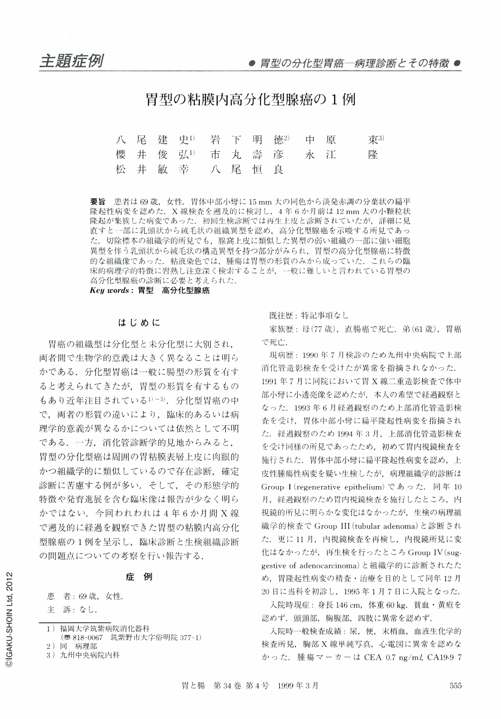
要旨 患者は69歳,女性.胃体中部小彎に15mm大の同色から淡発赤調の分葉状の扁平隆起性病変を認めた.X線検査を遡及的に検討し,4年6か月前は12mm大の小顆粒状隆起が集簇した病変であった.初回生検診断では再生上皮と診断されていたが,詳細に見直すと一部に乳頭状から絨毛状の組織異型を認め,高分化型腺癌を示唆する所見であった.切除標本の組織学的所見でも,腺窩上皮に類似した異型の弱い組織の一部に強い細胞異型を伴う乳頭状から絨毛状の構造異型を持つ部分がみられ,胃型の高分化型腺癌に特徴的な組織像であった.粘液染色では,腫瘍は胃型の形質のみから成っていた.これらの臨床的病理学的特徴に習熟し注意深く検索することが,一般に難しいと言われている胃型の高分化型腺癌の診断に必要と考えられた.
We reported a well differentiated adenocarcinoma of the gastric-type. The radiological examination of the carcinoma showed a slightly elevated lesion with lobulated structure 15 mm in diameter at the lesser curvarture of the middle part of the gastric body. The endoscopic study revealed the slightly red color of the lesion was the same as the non-cancerous background mucosa. The previous double contrast x-ray film was reviewed retrospectively and the radiological finding of the carcinoma taken 4.5 years before the operation showed an aggregated lesion of fine granular elevation 12 mm in diameter. The biopsy specimens orignally taken from this lesion had been diagnosed as benign regenerative epithelium. We reviewed these biopsy specimens histologically and found parts of the specimens showed severe cellular atypia with papillary and vinous structure, which is a characteristic finding of well differentiated adenocarcinoma of the gastric type. The histological investigation of the resected specimen showed the carcinoma was mainly composed of an atypical gland with very mild cellular and structural atypia and parts of it contained papillary and vinous glands with severe cellular atypia. By mucin histochemistry and immunohistochemistry staining, the carcinomatous tissue was shown to consist of only gastric type cells. Some difficulties were proposed in the clinical and pathological diagnosis of gastric-type carcinoma with well differentiation, because such a lesion resembled the non-cancerous surrounding foveolar epithelium. To avoid a misdiagnosis, we should understand the precise macroscopic and histological findings of well differentiated adenocarcinoma of the gastric-type.
Copyright © 1999, Igaku-Shoin Ltd. All rights reserved.