Japanese
English
- 有料閲覧
- Abstract 文献概要
- 1ページ目 Look Inside
- サイト内被引用 Cited by
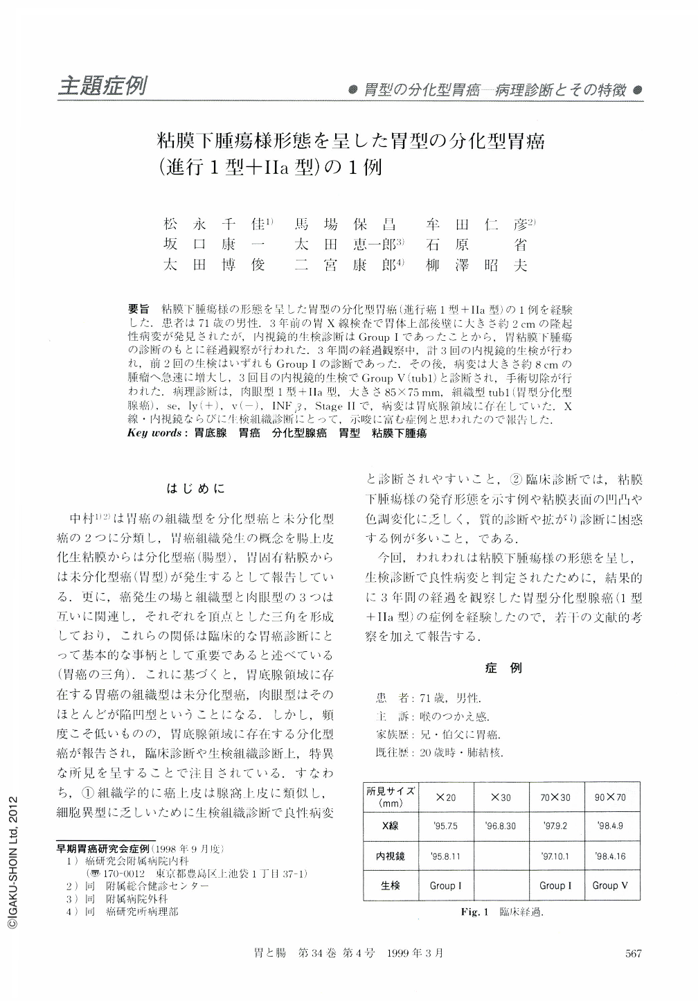
要旨 粘膜下腫瘍様の形態を呈した胃型の分化型胃癌(進行癌1型+Ⅱa型)の1例を経験した.患者は71歳の男性.3年前の胃X線検査で胃体上部後壁に大きさ約2cmの隆起性病変が発見されたが,内視鏡的生検診断はGroupⅠであったことから,胃粘膜下腫瘍の診断のもとに経過観察が行われた.3年間の経過観察中,計3回の内視鏡的生検が行われ,前2回の生検はいずれもGroupⅠの診断であった.その後,病変は大きさ約8cmの腫瘤へ急速に増大し,3回目の内視鏡的生検でGroupⅤ(tub1)と診断され,手術切除が行われた.病理診断は,肉眼型1型+Ⅱa型,大きさ85×75mm,組織型tub1(胃型分化型腺癌),se,ly(+),v(-),INFβ,StageⅡで,病変は胃底腺領域に存在していた.X線・内視鏡ならびに生検組織診断にとって,示唆に富む症例と思われたので報告した.
We report a case of differentiated adenocarcinoma of the stomach (advanced type Ⅰ and Ⅱa) mimicking a submucosal tumor. A 71-year-old man underwent upper gastrointestinal radiologic examination three years before, which detected an about 2 cm sized protruding tumor at the posterior wall of the upper body. Subsequent endoscopic biopsy examination showed group Ⅰ. During the follow-up period of three years, three endoscopic biopsies were performed and first two biopsy specimen were diagnosed as group Ⅰ, but the lesion grew rapidly about 8 cm in size and the third biopsy specimen revealed group Ⅴ (tub1). He underwent surgical resection. Pathologic diagnosis was macroscopically type Ⅰ + Ⅱa, 75 mm × 85 mm in size on the fundic gland area, and microscopically tub1 (differentiated adenocarcinoma of the stomach type), se, ly (+), v (-), INFβ, Stage Ⅱ. This case is, we believe, informative to the radiologic, endoscopic and biopsy diagnosis.

Copyright © 1999, Igaku-Shoin Ltd. All rights reserved.


