Japanese
English
- 有料閲覧
- Abstract 文献概要
- 1ページ目 Look Inside
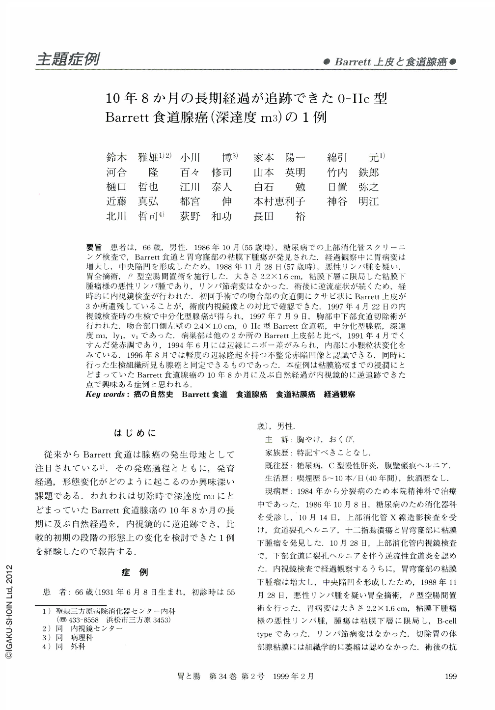
要旨 患者は,66歳,男性.1986年10月(55歳時),糖尿病での上部消化管スクリーニング検査で,Barrett食道と胃穹窿部の粘膜下腫瘍が発見された.経過観察中に胃病変は増大し,中央陥凹を形成したため,1988年11月28日(57歳時),悪性リンパ腫を疑い,胃全摘術,ρ型空腸間置術を施行した.大きさ2.2×1.6cm,粘膜下層に限局した粘膜下腫瘤様の悪性リンパ腫であり,リンパ節病変はなかった.術後に逆流症状が続くため,経時的に内視鏡検査が行われた.初回手術での吻合部の食道側にクサビ状にBarrett上皮が3か所遺残していることが,術前内視鏡像との対比で確認できた.1997年4月22日の内視鏡検査時の生検で中分化型腺癌が得られ,1997年7月9日,胸部中下部食道切除術が行われた.吻合部口側左壁の2.4×1.0cm,0-Ⅱc型Barrett食道癌,中分化型腺癌,深達度m3,ly1,v1であった.病巣部は他の2か所のBarrett上皮部と比べ,1991年4月でくすんだ発赤調であり,1994年6月には辺縁にニボー差がみられ,内部に小顆粒状変化をみている.1996年8月では軽度の辺縁隆起を持つ不整発赤陥凹像と認識できる.同時に行った生検組織所見も腺癌と同定できるものであった.本症例は粘膜筋板までの浸潤にとどまっていたBarrett食道腺癌の10年8か月に及ぶ自然経過が内視鏡的に逆追跡できた点で興味ある症例と思われる.
The patient is a 66-year-old male now. He visited our department because of diabetes mellitus in October, 1986 (at 55 years of age). On upper GI study, Barrett's esophagus and a small submucosal tumor in the gastric fornix was detected during a follow-up period of 25 months, the gastric tumor increased in size, and developed a central depression, suggestive of malignant lymphoma. On November 28, 1988 (at 57 years of age), total gastrectomy and jejunum interportion (ρ) was performed. Pathologically, the gastric tumor was diagnosed as B-cell type malignant lymphoma, 2.2 × 1.6 cm in size, limted to the submucosal layer. It looked like a submucosal tumor and there was no lympho node metastasis. Because of gastro-esophageal reflex syndrome after the operation, follow-up endoscopic examination was carried out. Three wedge-shaped remnants of Barrett's epithelium at the oral side esophagus of the anastomosis were confirmed and were compared with a preoperative endoscopic picture. Pathological stadies on an endoscopical forcep biopsy specimen on April 22, 1997 revealed a moderately differentiated adnocarcinoma, so middle, lower thoracic esophagectomy was peformed on July 9, 1997. Pathological studies on the resected specimen revealed type 0-Ⅱc Barrett's esophageal carcinoma on the left wall oral side of the anastomosis, 2.4 × 1.0 cm in size. It was a moderately differentiated adenocarcinoma, with a depth of invasion of m3, ly1, v1. In contrast with another two cases of Barrett's epithelium, the lesion was dull reddish in color in April, 1991 and had developed a slightly depressed configuration with fine granular change in June, 1994. In Angust, 1996, the lesion was recognized as having an irregalar reddish depression nith slight marginal elevation. Retrospectively, the same time endoscopic forcep biopsy specimen taken at this time was suggesive of adenocarcinoma.
We would like to emphasize the usefulness and importance of periodical endoscopic and biopsy follow-up for Barrett's esophagus.

Copyright © 1999, Igaku-Shoin Ltd. All rights reserved.


