Japanese
English
- 有料閲覧
PPV購入案内
1,320円 (1,200円+税10%) こちらは、91ページから98ページの文献です。購入には医書.jp本会員登録が必要です。
会員登録完了後に改めて上記「購入サイトへ」を選択してください。
または「購入サイトへ」選択後に「購入ログイン」、「新規会員登録」の順に進んでください。
会員登録について詳しくはをご確認ください。
- Abstract 文献概要
- 1ページ目 Look Inside
- サイト内被引用 Cited by
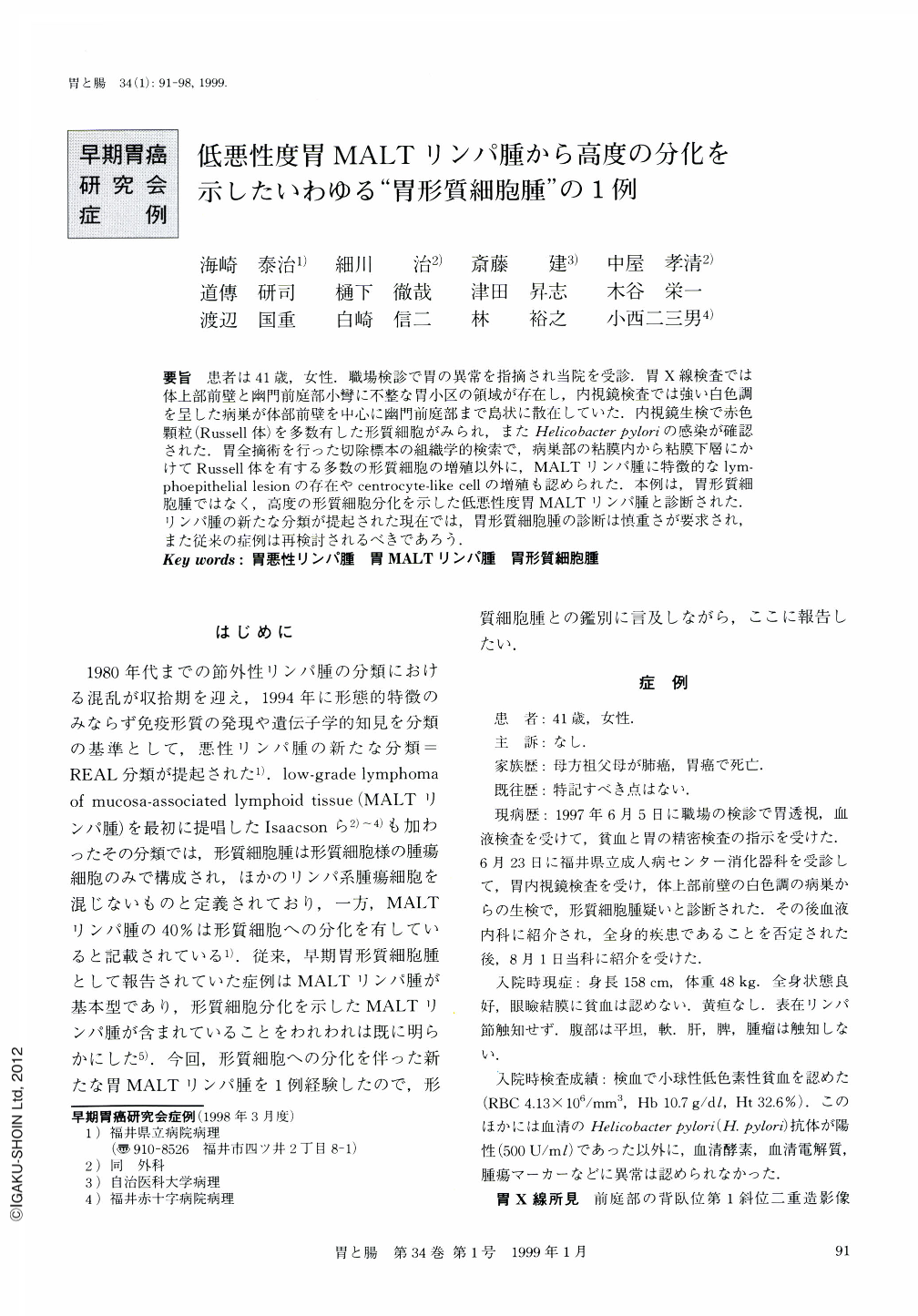
要旨 患者は41歳,女性.職場検診で胃の異常を指摘され当院を受診.胃X線検査では体上部前壁と幽門前庭部小彎に不整な胃小区の領域が存在し,内視鏡検査では強い白色調を呈した病巣が体部前壁を中心に幽門前庭部まで島状に散在していた.内視鏡生検で赤色顆粒(Russell体)を多数有した形質細胞がみられ,またHelicobacter pyloriの感染が確認された.胃全摘術を行った切除標本の組織学的検索で,病巣部の粘膜内から粘膜下層にかけてRussell体を有する多数の形質細胞の増殖以外に,MALTリンパ腫に特徴的なlymphoepithelial lesionの存在やcentrocyte-like cellの増殖も認められた.本例は,胃形質細胞腫ではなく,高度の形質細胞分化を示した低悪性度胃MALTリンパ腫と診断された.リンパ腫の新たな分類が提起された現在では,胃形質細胞腫の診断は慎重さが要求され,また従来の症例は再検討されるべきであろう.
A case of low-grade B-cell gastric lymphoma of mucosa-associated lymphoid tissue (MALT) exhibiting marked plasma cell differentiation is reported. A 41-year-old female patient was referred to the Fukui Prefectural Hospital because of some abnormality of the stomach indicated at a medical examination. X-ray and endoscopic examination revealed white irregular granular mucosa in the upper body and antrum of the stomach. Pathological examination of the biopsy specimen revealed diffuse plasma cell infiltration with many red crystalline inclusions and Helicobacter pylori infection. The patient received total gastrectomy. Histologically, intense infiltration of centrocyte-like (CCL) cells was observed spread in the mucosa. These CCL cells formed lymphoepithelial lesions in the mucosa. Besides these findings, CCL cells, with red crystalline inclusions, were clearly differentiated from plasma cells. Considering these points, a difinite pathologic diagnosis of low-grade B-cell gastric lymphoma of MALT with marked plasma cell differentiation was made. As the REAL classification of malignant lymphoma has been newly adopted, the diagnosis of gastric“plasmacytoma” should be made carefully, and we should reconsider the cases formerly diagnosed as gastric“plasmacytoma”.
Copyright © 1999, Igaku-Shoin Ltd. All rights reserved.