Japanese
English
- 有料閲覧
PPV購入案内
1,320円 (1,200円+税10%) こちらは、1750ページから1756ページの文献です。購入には医書.jp本会員登録が必要です。
会員登録完了後に改めて上記「購入サイトへ」を選択してください。
または「購入サイトへ」選択後に「購入ログイン」、「新規会員登録」の順に進んでください。
会員登録について詳しくはをご確認ください。
- Abstract 文献概要
- 1ページ目 Look Inside
- 参考文献 Reference
- サイト内被引用 Cited by
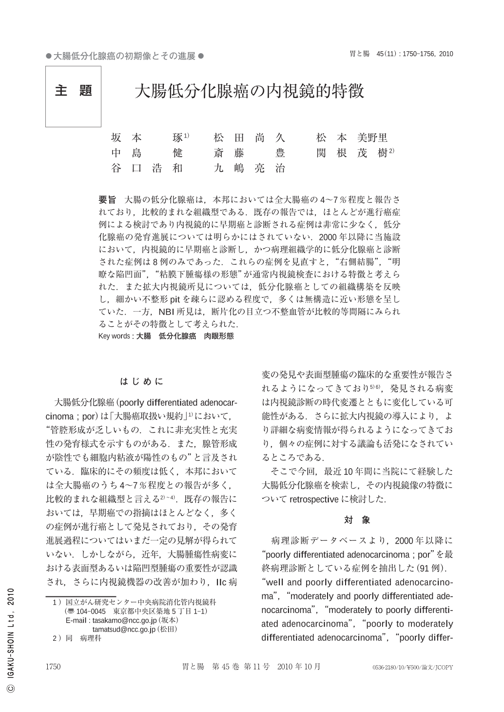
要旨 大腸の低分化腺癌は,本邦においては全大腸癌の4~7%程度と報告されており,比較的まれな組織型である.既存の報告では,ほとんどが進行癌症例による検討であり内視鏡的に早期癌と診断される症例は非常に少なく,低分化腺癌の発育進展については明らかにはされていない.2000年以降に当施設において,内視鏡的に早期癌と診断し,かつ病理組織学的に低分化腺癌と診断された症例は8例のみであった.これらの症例を見直すと,“右側結腸",“明瞭な陥凹面",“粘膜下腫瘍様の形態"が通常内視鏡検査における特徴と考えられた.また拡大内視鏡所見については,低分化腺癌としての組織構築を反映し,細かい不整形pitを疎らに認める程度で,多くは無構造に近い形態を呈していた.一方,NBI所見は,断片化の目立つ不整血管が比較的等間隔にみられることがその特徴として考えられた.
Poorly differentiated adenocarcinoma is considered to be a relatively uncommon histological type in the colorectum. Its origin or progression is still unclear, because most of the reports have been mainly based on clinicopathological features of advanced colorectal cancer. Relatively little has been learned about it in the early stage, or about lesions showing early-stage configuration endoscopically. In all patients who underwent endoscopic or surgical treatment between 2000 and 2009 in the National Cancer Center Hospital, Tokyo, there have been 34 cases diagnosed as having poorly differentiated adenocarcinoma as the predominant histological component. Among them, there were 8 cases judged as early stage cancer. Reviewing these cases, two gross findings appeared to be common findings : “clearly demarcated depressed area" and “submucosal-tumor-like configuration". Moreover, most of them(7/8)were located in the right-sided colon. These clinical findings are probably important features of poorly differentiated adenocarcinoma of the colon. Regarding the findings of magnifying chromoendoscopy, a few small and fine pits were observed in the depressed area, and, using narrow band imaging, fragmented irregular vessels running in a relatively well-ordered pattern were observed. This may reflect the histological intramucosal structure of this tumor. It is difficult to make conclusive remarks on the endoscopic finding especially magnifying chromoendoscopy, as more detailed analysis using a large number of cases is required.
Copyright © 2010, Igaku-Shoin Ltd. All rights reserved.