Japanese
English
- 有料閲覧
- Abstract 文献概要
- 1ページ目 Look Inside
- 参考文献 Reference
- サイト内被引用 Cited by
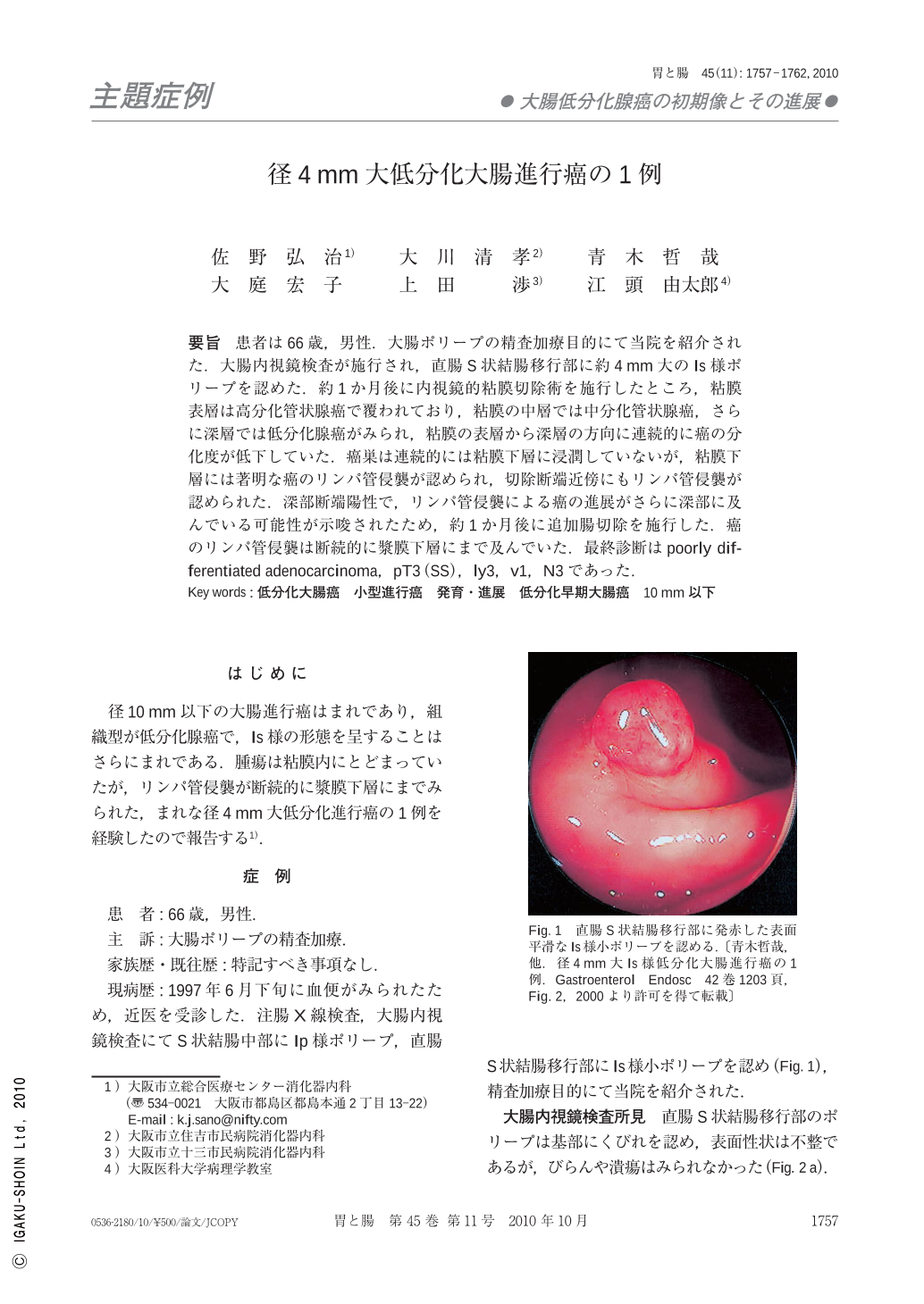
要旨 患者は66歳,男性.大腸ポリープの精査加療目的にて当院を紹介された.大腸内視鏡検査が施行され,直腸S状結腸移行部に約4mm大のIs様ポリープを認めた.約1か月後に内視鏡的粘膜切除術を施行したところ,粘膜表層は高分化管状腺癌で覆われており,粘膜の中層では中分化管状腺癌,さらに深層では低分化腺癌がみられ,粘膜の表層から深層の方向に連続的に癌の分化度が低下していた.癌巣は連続的には粘膜下層に浸潤していないが,粘膜下層には著明な癌のリンパ管侵襲が認められ,切除断端近傍にもリンパ管侵襲が認められた.深部断端陽性で,リンパ管侵襲による癌の進展がさらに深部に及んでいる可能性が示唆されたため,約1か月後に追加腸切除を施行した.癌のリンパ管侵襲は断続的に漿膜下層にまで及んでいた.最終診断はpoorly differentiated adenocarcinoma,pT3(SS),ly3,v1,N3であった.
A 66-year-old man was introduced to this hospital for further examination and treatment of polyps in the colon. An Is type polyp(4mm in diameter)was observed in the recto-sigmoid colon by colonoscopy. Endoscopic mucosal resection was performed, and poorly differentiated adenocarcinoma in the bottom layer mixed with well differentiated tubular adenocarcinoma in the surface layer and moderately differentiated adenocarcinoma in the middle layer was found in the mucosal layer. Although the tumor did not pass through the muscular layer of the mucosa, it infiltrated into the submucosal lymphatic vessel, so an additional operation was conducted. Vascular invasion had spread to the lymphatic vessels in the surrounding adipose tissue. The illness was diagnosed as a poorly differentiated adenocarcinoma such as pT3(ss), ly3, v1, N3.

Copyright © 2010, Igaku-Shoin Ltd. All rights reserved.


