Japanese
English
- 有料閲覧
PPV購入案内
1,320円 (1,200円+税10%) こちらは、335ページから344ページの文献です。購入には医書.jp本会員登録が必要です。
会員登録完了後に改めて上記「購入サイトへ」を選択してください。
または「購入サイトへ」選択後に「購入ログイン」、「新規会員登録」の順に進んでください。
会員登録について詳しくはをご確認ください。
- Abstract 文献概要
- 1ページ目 Look Inside
- 参考文献 Reference
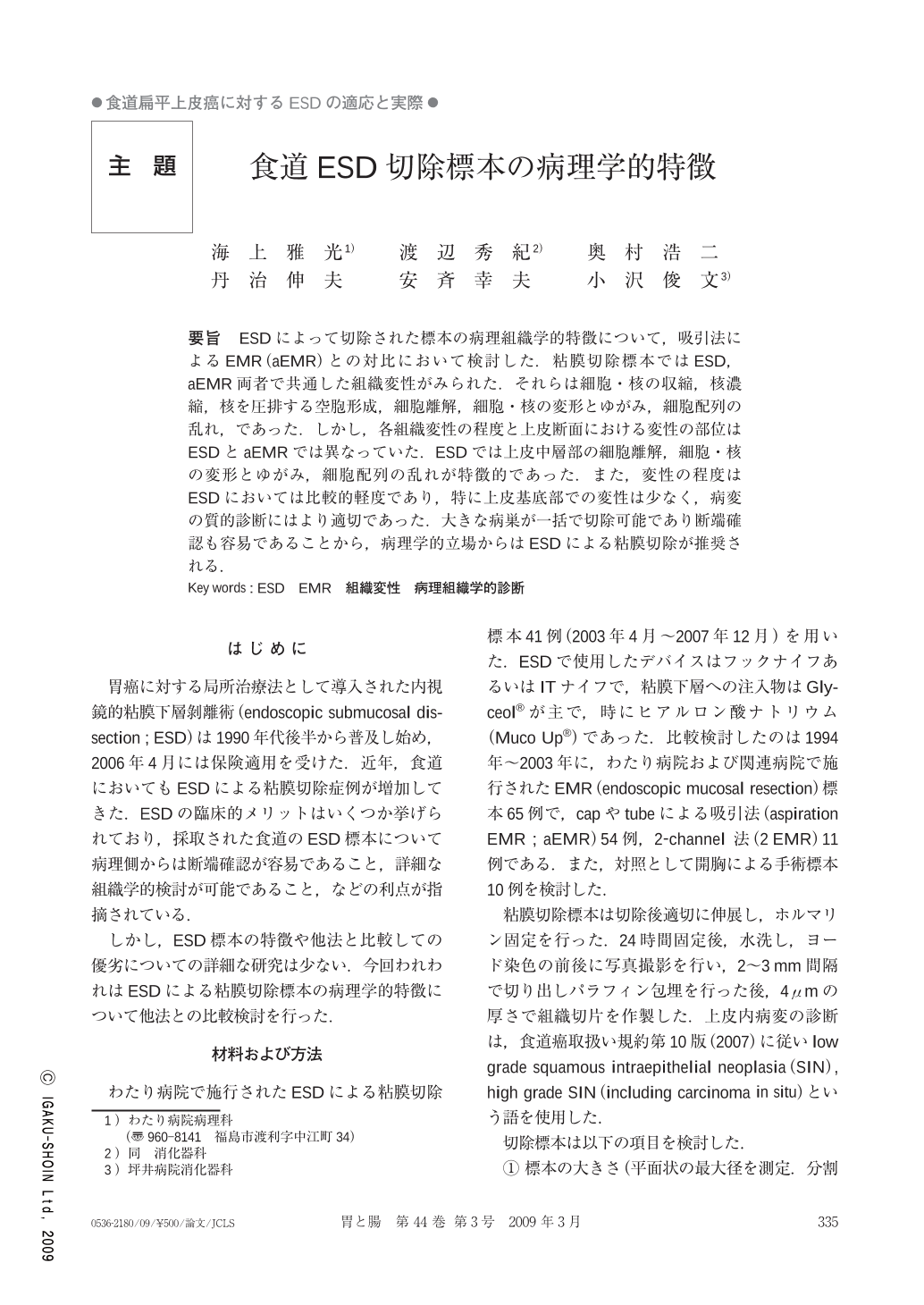
要旨 ESDによって切除された標本の病理組織学的特徴について,吸引法によるEMR(aEMR)との対比において検討した.粘膜切除標本ではESD,aEMR両者で共通した組織変性がみられた.それらは細胞・核の収縮,核濃縮,核を圧排する空胞形成,細胞離解,細胞・核の変形とゆがみ,細胞配列の乱れ,であった.しかし,各組織変性の程度と上皮断面における変性の部位はESDとaEMRでは異なっていた.ESDでは上皮中層部の細胞離解,細胞・核の変形とゆがみ,細胞配列の乱れが特徴的であった.また,変性の程度はESDにおいては比較的軽度であり,特に上皮基底部での変性は少なく,病変の質的診断にはより適切であった.大きな病巣が一括で切除可能であり断端確認も容易であることから,病理学的立場からはESDによる粘膜切除が推奨される.
We investigated the pathological characteristics of specimens obtained by ESD in comparison with those obtained by EMR. Several factors of degeneration were recognized as common to both ESD and EMR. They were shrinkage of cells and nuclei, pyknosis, perinuclear vacuolation, acantholysis, distortion of cells and nuclei, and disordered cell arrangement. In particular, specimens harvested by ESD showed acantholysis, distortion of cells and nuclei, and disordered cell arrangement. The degree of degeneration in ESD was slighter than that in EMR. Moreover degeneration in ESD was mainly observed in the middle layer of the epithelium and was relatively slight in the deep layer. These results revealed that specimens obtained by ESD were more suitable for the diagnosis of squamous intraepithelial neoplasia.
Copyright © 2009, Igaku-Shoin Ltd. All rights reserved.