Japanese
English
- 有料閲覧
- Abstract 文献概要
- 1ページ目 Look Inside
- サイト内被引用 Cited by
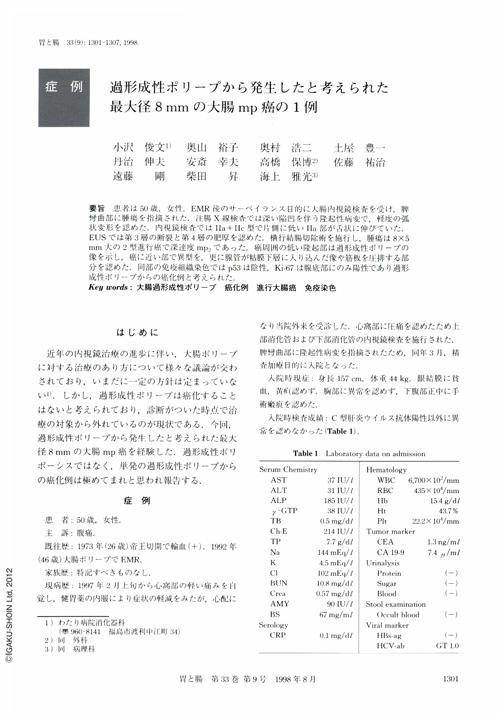
要旨 患者は50歳,女性.EMR後のサーベイランス目的に大腸内視鏡検査を受け,脾彎曲部に腫瘍を指摘された.注腸X線検査では深い陥凹を伴う隆起性病変で,軽度の弧状変形を認めた.内視鏡検査ではⅡa+Ⅱc型で片側に低いⅡa部が舌状に伸びていた.EUSでは第3層の断裂と第4層の肥厚を認めた.横行結腸切除術を施行し,腫瘍は8×5mm大の2型進行癌で深達度mp2であった.癌周囲の低い隆起部は過形成性ポリープの像を示し,癌に近い部で異型を,更に腺管が粘膜下層に入り込んだ像や筋板を圧排する部分を認めた.同部の免疫組織染色ではp53は陰性,Ki-67は腺底部にのみ陽性であり過形成性ポリープからの癌化例と考えられた.
A 50-year-old female who had a history of mucosectomy of a colonic polyp four years previously was admitted to our hospital for surveillance colonoscopy. Ⅱa+Ⅱc-like lesion associated with broad and flat elevation was detected at the splenic flexure where a deformity of the colonic wall was also noticed. EUS (20 MHz) examination showed the destroyed third layer and thickening of the fourth layer. Endoscopic diagnosis was type Ⅱa+Ⅱc colonic cancer and cancer invasion was estimated as sm3~mp. Partial colectomy with lymph node dissection was performed. Pathological examination revealed that the lesion consisted of a cancerous area and a hyperplastic polyp. The cancer was a papillary adenocarcinoma, 8×4 mm in size and depth of invasion was to the mp. The adenocarcinoma was surrounded by a hyperplastic polyp which was 20×5 mm in size. In the hyperplastic polyp, Ki-67 was positive at the lower part of the gland. Adenocarcinoma and hyperplastic polyp were negative for p53 staining. The lesion was thought to be advanced cancer originating from a solitary hyperplastic polyp.

Copyright © 1998, Igaku-Shoin Ltd. All rights reserved.


