Japanese
English
- 有料閲覧
PPV購入案内
1,320円 (1,200円+税10%) こちらは、1283ページから1289ページの文献です。購入には医書.jp本会員登録が必要です。
会員登録完了後に改めて上記「購入サイトへ」を選択してください。
または「購入サイトへ」選択後に「購入ログイン」、「新規会員登録」の順に進んでください。
会員登録について詳しくはをご確認ください。
- Abstract 文献概要
- 1ページ目 Look Inside
- 参考文献 Reference
- サイト内被引用 Cited by
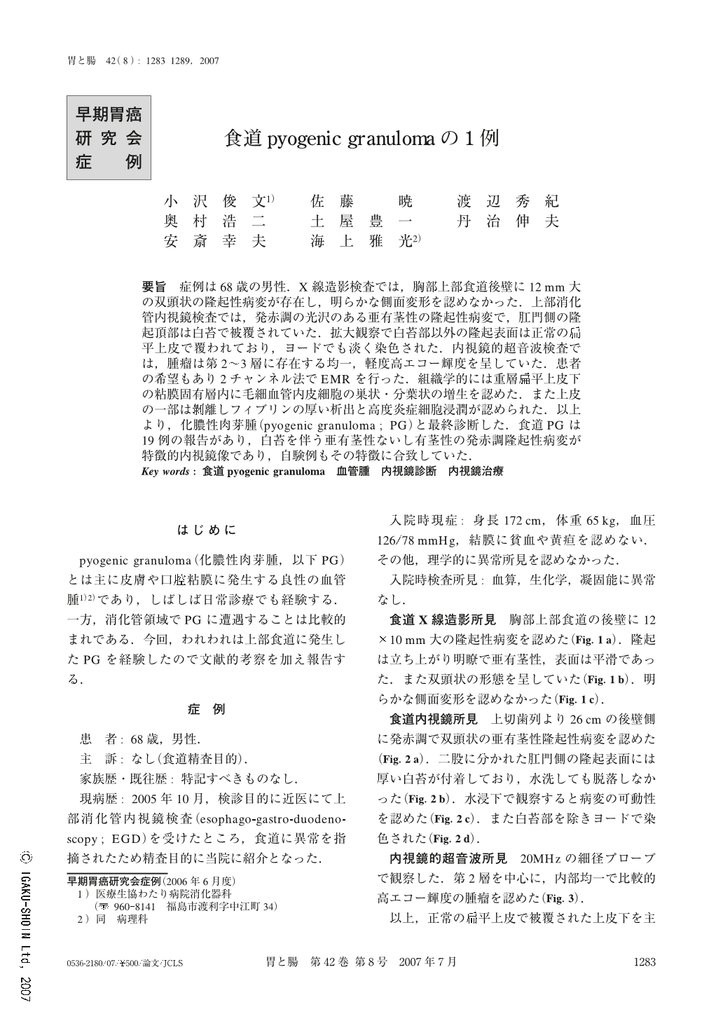
要旨 症例は68歳の男性.X線造影検査では,胸部上部食道後壁に12mm大の双頭状の隆起性病変が存在し,明らかな側面変形を認めなかった.上部消化管内視鏡検査では,発赤調の光沢のある亜有茎性の隆起性病変で,肛門側の隆起頂部は白苔で被覆されていた.拡大観察で白苔部以外の隆起表面は正常の扁平上皮で覆われており,ヨードでも淡く染色された.内視鏡的超音波検査では,腫瘤は第2~3層に存在する均一,軽度高エコー輝度を呈していた.患者の希望もあり2チャンネル法でEMRを行った.組織学的には重層扁平上皮下の粘膜固有層内に毛細血管内皮細胞の巣状・分葉状の増生を認めた.また上皮の一部は剥離しフィブリンの厚い析出と高度炎症細胞浸潤が認められた.以上より,化膿性肉芽腫(pyogenic granuloma;PG)と最終診断した.食道PGは19例の報告があり,白苔を伴う亜有茎性ないし有茎性の発赤調隆起性病変が特徴的内視鏡像であり,自験例もその特徴に合致していた.
Pyogenic granuloma of the esophagus is rare, and only 18 cases of this disease have been reported. We reported a 19th case of pyogenic granuloma of the esophagus in Japan.
A 68-year-old man requesting a medical check was admitted to our hospital. Esophagography demonstrated a double-headed protrusion, which was about 12mm in size, with no profile deformation, in the upper esophagus. Endoscopic examination revealed a pedunculated polypoid lesion with redness which was covered with a white coating on the top. Magnifying endoscopy showed a normal squamous epithelium with iodine staining on the surface of the protrusion. EUS revealed a slightly high echoic mass in the 2nd to 3rd layer of the esophageal wall. EMR was performed using a double-channel scope and no complications occurred. Histological examination of the resected tumor revealed subepithelial proliferation of capillary vessels with infiltration of inflammatory cells. White coating on the lesion was composed of a thick layer of fibrin. For this reason, diagnosis of pyogenic granuloma was made. Characteristic endoscopic findings of pyogenic granuloma in the esophagus were a pedunculated polypoid lesion having a reddish stalk and which was covered with a white coating on its top.
Copyright © 2007, Igaku-Shoin Ltd. All rights reserved.