Japanese
English
- 有料閲覧
PPV購入案内
1,320円 (1,200円+税10%) こちらは、248ページから257ページの文献です。購入には医書.jp本会員登録が必要です。
会員登録完了後に改めて上記「購入サイトへ」を選択してください。
または「購入サイトへ」選択後に「購入ログイン」、「新規会員登録」の順に進んでください。
会員登録について詳しくはをご確認ください。
- Abstract 文献概要
- 1ページ目 Look Inside
- 参考文献 Reference
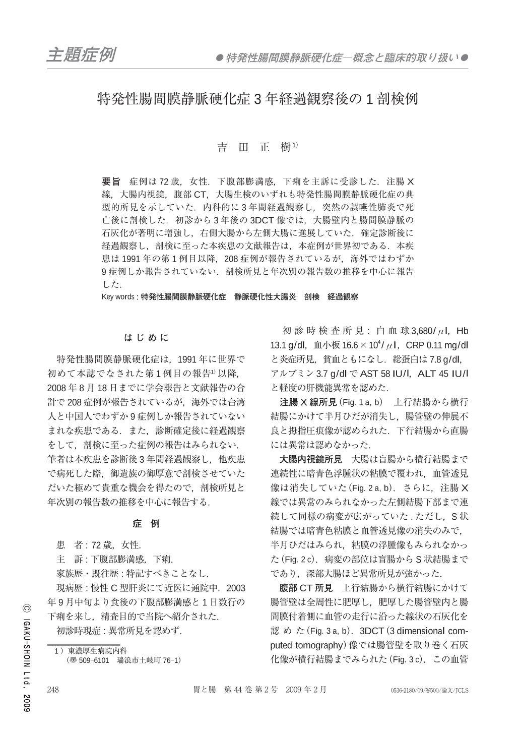
要旨 症例は72歳,女性.下腹部膨満感,下痢を主訴に受診した.注腸X線,大腸内視鏡,腹部CT,大腸生検のいずれも特発性腸間膜静脈硬化症の典型的所見を示していた.内科的に3年間経過観察し,突然の誤嚥性肺炎で死亡後に剖検した.初診から3年後の3DCT像では,大腸壁内と腸間膜静脈の石灰化が著明に増強し,右側大腸から左側大腸に進展していた.確定診断後に経過観察し,剖検に至った本疾患の文献報告は,本症例が世界初である.本疾患は1991年の第1例目以降,208症例が報告されているが,海外ではわずか9症例しか報告されていない.剖検所見と年次別の報告数の推移を中心に報告した.
Idiopathic mesenteric phlebosclerosis is a recently discovered, rare and new disease entity causing chronic ischemic colitis. 208 cases of this disease have been reported since 1991. Japanese cases numbered 199, Taiwanese cases numbered 8 and there was only one Chinese case. However, this disease has not been reported among other races. This is a first autopsy case after a 3- year period of observation. A 72- year-old woman visited our hospital because of lower abdominal fullness and slight diarrhea. Barium enema showed the disappearance of semilunar folds and the thumbprinting appearance of the ascending colon and transverse colon. On the other hand, almost normal findings were obtained in the left hemicolon. Colonoscopic examination showed dark purple and edematous mucosa with loss of the normal vascular pattern in the ascending colon and transverse colon. In the sigmoid colon, colonoscopic examination showed only dark purple mucosa without mucosal edema and disappearance of semilunar folds. Abdominal CT showed bowel wall thickening with intramural calcifications in the cecum, ascending colon and transverse colon. 3DCT showed calcifications around the right hemicolon. After 3 years, calcifications around the colon and along the mesentery markedly increased and extended to the left hemicolon. The patient suddenly died because of severe aspiration pneumonia. An autopsy was carried out. Macroscopic findings of the resected specimen showed dark purple thickened ascending colon and transverse colon. Ileum and appendix were normal. A radiograph of the resected colon showed tortuous threadlike calcifications within the cecum, ascending colon, transverse colon and descending colon. These calcifications predominantly affected the right hemicolon. Microscopic examination showed marked fibrous thickening of the submucosal layer. The veins in the colonic wall showed tortuous appearance. Marked fibrous thickening of the venous walls with calcification, ossification and recanalization showed severe luminal narrowing. Perivascular fibrosis of the intramural colonic veins was also found. These findings were consistent with mesenteric phlebosclerosis.
Copyright © 2009, Igaku-Shoin Ltd. All rights reserved.