Japanese
English
- 有料閲覧
- Abstract 文献概要
- 1ページ目 Look Inside
- 参考文献 Reference
- サイト内被引用 Cited by
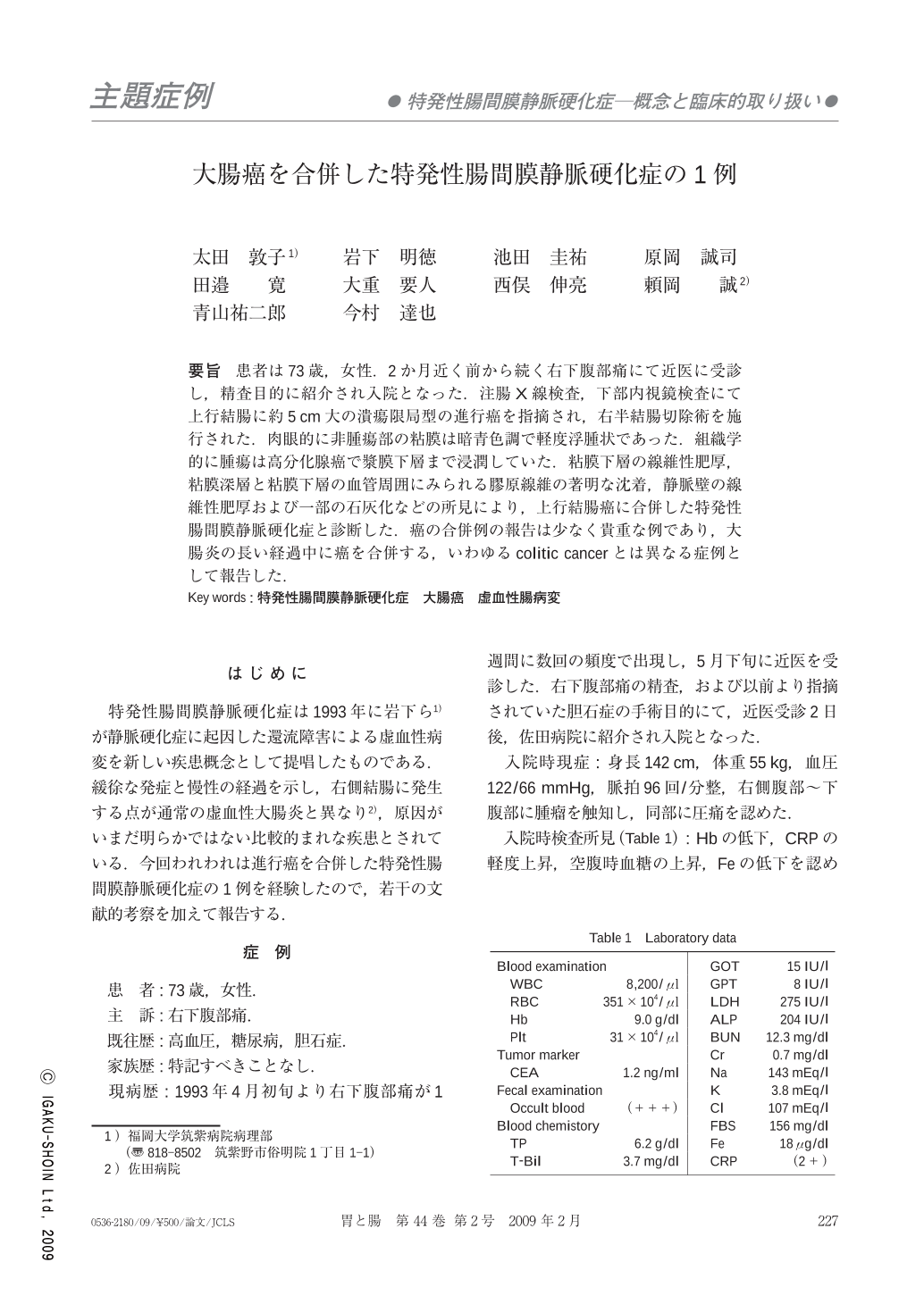
要旨 患者は73歳,女性.2か月近く前から続く右下腹部痛にて近医に受診し,精査目的に紹介され入院となった.注腸X線検査,下部内視鏡検査にて上行結腸に約5cm大の潰瘍限局型の進行癌を指摘され,右半結腸切除術を施行された.肉眼的に非腫瘍部の粘膜は暗青色調で軽度浮腫状であった.組織学的に腫瘍は高分化腺癌で漿膜下層まで浸潤していた.粘膜下層の線維性肥厚,粘膜深層と粘膜下層の血管周囲にみられる膠原線維の著明な沈着,静脈壁の線維性肥厚および一部の石灰化などの所見により,上行結腸癌に合併した特発性腸間膜静脈硬化症と診断した.癌の合併例の報告は少なく貴重な例であり,大腸炎の長い経過中に癌を合併する,いわゆるcolitic cancerとは異なる症例として報告した.
A 73-year-old woman was admitted to the hospital with the complaint of right lower quadrant(RLQ)pain for about two months. Barium enema examination and colonoscopy revealed an ascending colon adenocarcinoma. A right hemicolectomy was performed. Macroscopically, the carcinoma was 5.5×5.0cm in size, and the large intestine showed a slightly dark purple-colored surface, swelling, and mild thickening of the wall. Pathologically, the tumor was a T3N0 well differentiated adenocarcioma. Sections also showed edema, veins with fibrous thickening and mild fibrosis in the submucosa, and deposition of collagen around vessels in the mucosa. Only a few serosal veins showed mural calcification. The marked thickening submucosal layer was mainly because of edema and mild fibrosis. The histological findings were consistent with those of idiopathic mesenteric phlebosclerosis(IMP), and also suggested an early stage of this disease. We concluded that the carcinoma was coincident with IMP, and was not a so-called colitic cancer.

Copyright © 2009, Igaku-Shoin Ltd. All rights reserved.


