Japanese
English
- 有料閲覧
- Abstract 文献概要
- 1ページ目 Look Inside
- 参考文献 Reference
- サイト内被引用 Cited by
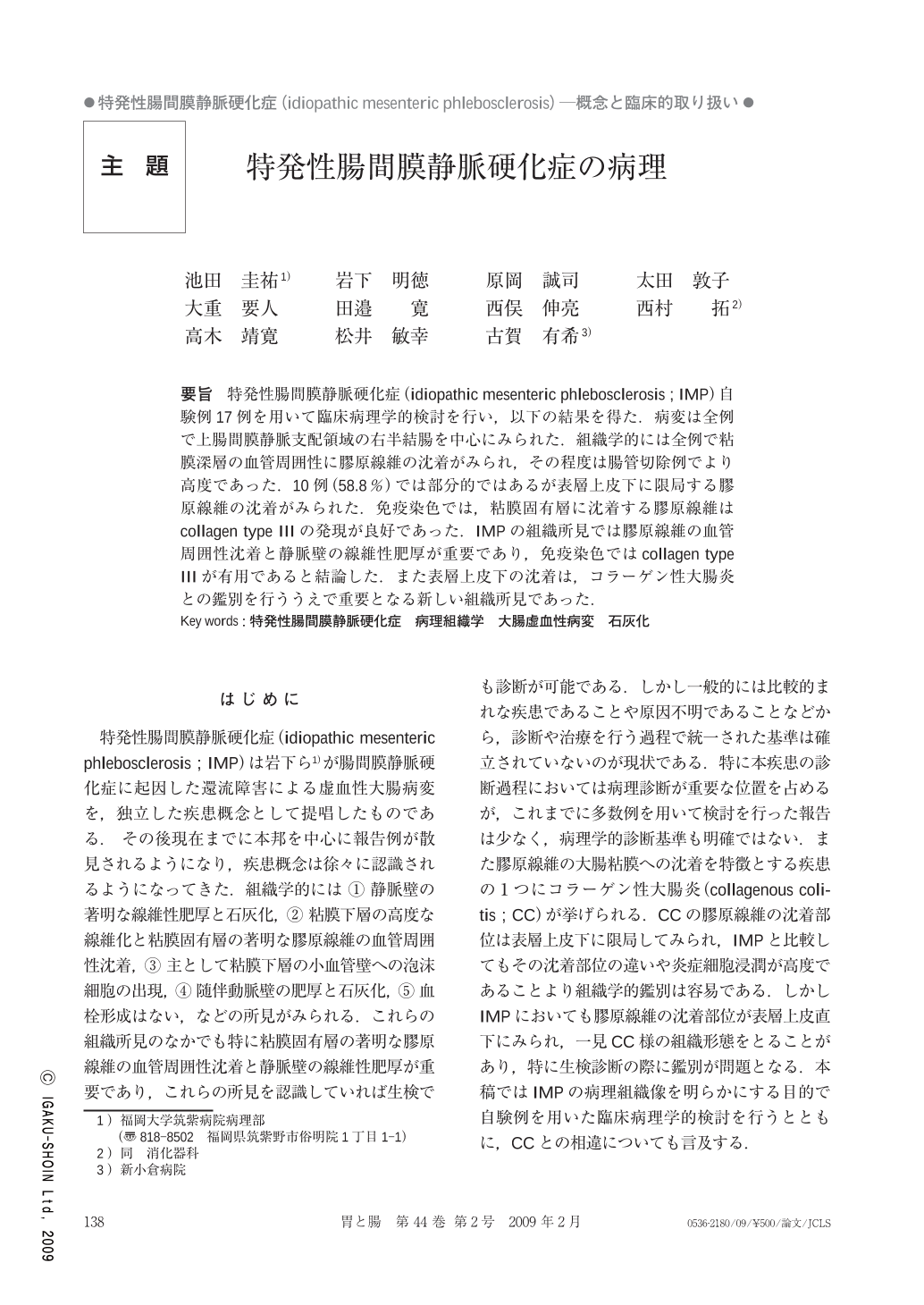
要旨 特発性腸間膜静脈硬化症(idiopathic mesenteric phlebosclerosis;IMP)自験例17例を用いて臨床病理学的検討を行い,以下の結果を得た.病変は全例で上腸間膜静脈支配領域の右半結腸を中心にみられた.組織学的には全例で粘膜深層の血管周囲性に膠原線維の沈着がみられ,その程度は腸管切除例でより高度であった.10例(58.8%)では部分的ではあるが表層上皮下に限局する膠原線維の沈着がみられた.免疫染色では,粘膜固有層に沈着する膠原線維はcollagen type Ⅲの発現が良好であった.IMPの組織所見では膠原線維の血管周囲性沈着と静脈壁の線維性肥厚が重要であり,免疫染色ではcollagen type Ⅲが有用であると結論した.また表層上皮下の沈着は,コラーゲン性大腸炎との鑑別を行ううえで重要となる新しい組織所見であった.
We investigated the clinical pathology in 17 cases of idiopathic mesenteric phlebosclerosis(IMP)at our hospital. All lesions were located mainly on the right side of the colon in the upper mesenteric vein area. Histopathological examinations revealed collagen fiber deposits around vessels in the deep mucosa in all cases, which were more severe in the cases treated with intestinal resection. Ten cases(58.8%)showed focal collagen fiber deposits in the superficial subepithelial tissue. Immunostaining studies demonstrated rich collagen type Ⅲ expression in the collagen fibers in the lamina propria mucosa layer. We concluded that collagen fiber deposits around vessels are a characteristic finding in the histopathology of IMP, and that detection of collagen type Ⅲ expression by immunostaining is a feasible diagnostic method. Moreover, the presence of deposits in the superficial subepithelial tissue is a new significant histological finding that will enable IMP to be distinguished from collagenous colitis.

Copyright © 2009, Igaku-Shoin Ltd. All rights reserved.


