Japanese
English
- 有料閲覧
PPV購入案内
1,320円 (1,200円+税10%) こちらは、343ページから349ページの文献です。購入には医書.jp本会員登録が必要です。
会員登録完了後に改めて上記「購入サイトへ」を選択してください。
または「購入サイトへ」選択後に「購入ログイン」、「新規会員登録」の順に進んでください。
会員登録について詳しくはをご確認ください。
- Abstract 文献概要
- 1ページ目 Look Inside
- 参考文献 Reference
- サイト内被引用 Cited by
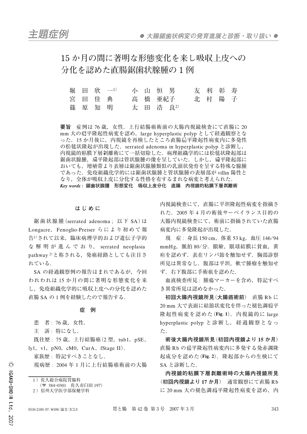
要旨 症例は76歳,女性.上行結腸癌術前の大腸内視鏡検査にて直腸に20mm大の扁平隆起性病変を認め,large hyperplastic polypとして経過観察となった.15か月後に,内視鏡を再検したところ直腸扁平隆起性病変内に多発性の松毬状隆起が出現した.serrated adenoma in hyperplastic polypと診断し,内視鏡的粘膜下層剥離術にて一括切除した.病理組織学的には松毬状隆起部は鋸歯状腺腫,扁平隆起部は管状腺腫の像を呈していた.しかし,扁平隆起部においても,増殖帯より表層は鋸歯状腺腫類似の乳頭状発育を呈する特殊な腺腫であった.免疫組織化学的には鋸歯状腺腫と管状腺腫の表層部がvillin陽性となり,全体が吸収上皮に分化する性格を有するまれな病変と考えられた.
A 76-year-old female underwent first colonoscopy before colectomy for advanced ascending colon cancer. A slightly elevated lesion, 20mm in size, was detected and diagnosed as a large hyperplastic polyp. She underwent a second colonoscopy 15 months later, multiple pine-cone like protrusions appeared in the slightly elevated lesion of the rectum. We diagnosed it as a serrated adenoma in a hyperplastic polyp, and an en-bloc resection was performed by endoscopic submucosal dissection. Histological diagnosis revealed that, pathologically, there was a mixture of two types of adenoma; serrated adenoma with protrusion and tubular adenoma with slight elevation. However, papillary growth pattern like serrated adenoma was seen at the surface of the tubular adenoma. Immnunohistochemical staining of villin was positive in the surface of both serrated adenoma and tubular adenoma, so we considered it as having the character of absorptive enterocyte differentiation.
Copyright © 2007, Igaku-Shoin Ltd. All rights reserved.