Japanese
English
- 有料閲覧
- Abstract 文献概要
- 1ページ目 Look Inside
- 参考文献 Reference
- サイト内被引用 Cited by
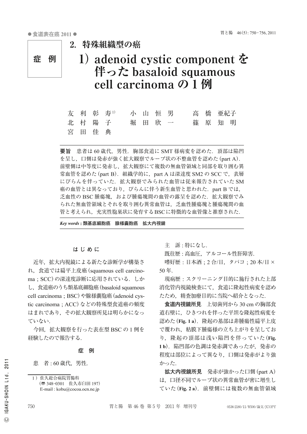
要旨 患者は60歳代,男性.胸部食道にSMT様病変を認めた.頂部は陥凹を呈し,口側は発赤が強く拡大観察でループ状の不整血管を認めた(part A).前壁側は中等度に発赤し,拡大観察にて複数の無血管領域と同部を取り囲む異常血管を認めた(part B).組織学的に,part Aは深達度SM2のSCCで,表層にびらんを伴っていた.拡大観察でみられた血管は従来報告されていたSM癌の血管とは異なっており,びらんに伴う新生血管と思われた.part Bでは,乏血性のBSC腫瘍塊,および腫瘍塊間の血管の露呈を認めた.拡大観察でみられた無血管領域とそれを取り囲む異常血管は,乏血性腫瘍塊と腫瘍塊間の血管と考えられ,充実性胞巣状に発育するBSCに特徴的な血管像と推察された.
A 60s-year-old male underwent esophago-gastroscopy. A flat elevated lesion with central depression was observed in the middle esophagus. The color of the oral part was red and magnifying endoscopy showed loop-shaped irregular vessels(part A). The color of the anterior part was moderate red and there were some depressed a-vascular areas with unusual surrounding vessels(part B).
Histologically, regenerative vessels caused by erosion were observed on the surface of part A, and SCC(squamous cell carcinoma)had invaded to SM2. The vessels of part A observed with magnifying endoscopy were different from those of the part where SCC invaded the submucosa. So, the loop-shaped vessels were considered to be regenerative vessels.
Microscopic findings of part B revealed the exposure of BSC(basaloid squamous cell carcinoma)nests with few vessels and large vessels between BSC nests. The avascular areas of part B observed with magnifying endoscopy corresponded to BSC nests and the unusual vessels surrounding the avascular areas corresponded to the vessels between BSC nests. Therefore, these findings with magnifying endoscopy were considered to be the characteristic findings of BSC.

Copyright © 2011, Igaku-Shoin Ltd. All rights reserved.


