Japanese
English
- 有料閲覧
PPV購入案内
1,320円 (1,200円+税10%) こちらは、961ページから971ページの文献です。購入には医書.jp本会員登録が必要です。
会員登録完了後に改めて上記「購入サイトへ」を選択してください。
または「購入サイトへ」選択後に「購入ログイン」、「新規会員登録」の順に進んでください。
会員登録について詳しくはをご確認ください。
- Abstract 文献概要
- 1ページ目 Look Inside
- 参考文献 Reference
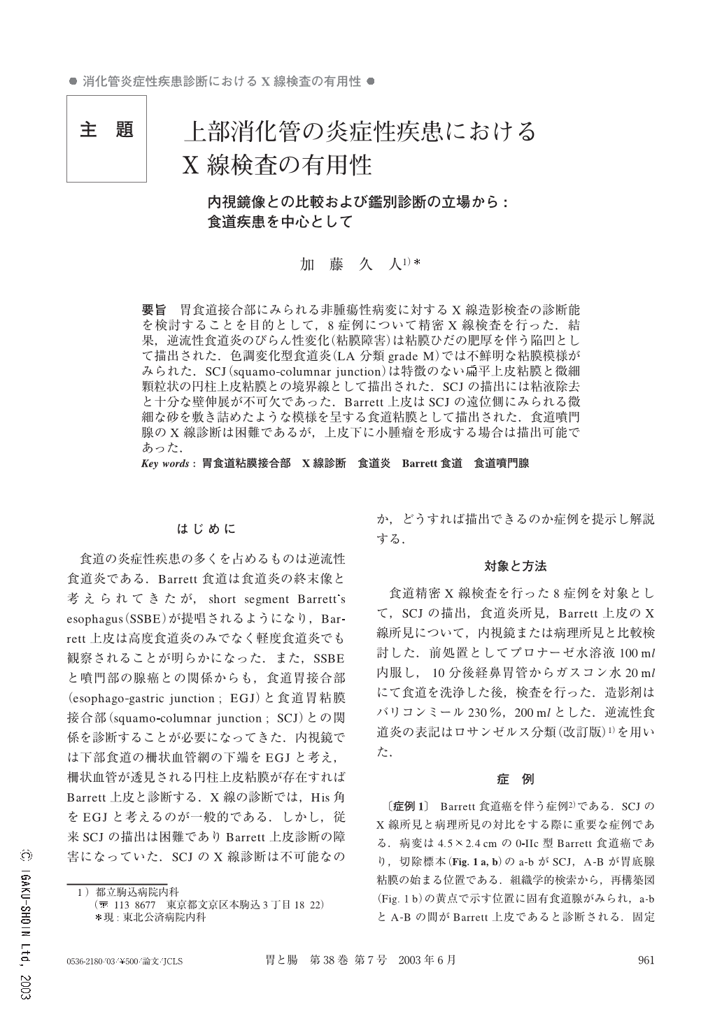
要旨 胃食道接合部にみられる非腫瘍性病変に対するX線造影検査の診断能を検討することを目的として,8症例について精密X線検査を行った.結果,逆流性食道炎のびらん性変化(粘膜障害)は粘膜ひだの肥厚を伴う陥凹として描出された.色調変化型食道炎(LA分類 grade M)では不鮮明な粘膜模様がみられた.SCJ(squamo-columnar junction)は特徴のない扁平上皮粘膜と微細顆粒状の円柱上皮粘膜との境界線として描出された.SCJの描出には粘液除去と十分な壁伸展が不可欠であった.Barrett上皮はSCJの遠位側にみられる微細な砂を敷き詰めたような模様を呈する食道粘膜として描出された.食道噴門腺のX線診断は困難であるが,上皮下に小腫瘤を形成する場合は描出可能であった.
The aim of this study was to investigate the ability of radiology for the diagnosis of non-cancerous lesions in the lower esophagus and in the esophagogastric junction. Eight patients were studied. One had normal SCJ (squamo-columnar junction), 2 had reflux esophagitis,3 had SSBE (short segment Barrett's esophagus), and the other three cases suffered from LSBE (long segment Barrett's esophagus), hiatal hernia, and esophageal cardial gland respectively.
SCJ was delineated in all cases, as the borderline between featureless squamous mucosa and small granular columnar epithelium. Mucosal breaks in reflux esophagitis were depicted as longitudinal depressions on the thickening folds. Discolored mucosa of grade M esophagitis was depicted as undulated surface mucosa. The features of Barrett's epithelium were seen as the fine granular surface mucosa adjacent to the SCJ.
Copyright © 2003, Igaku-Shoin Ltd. All rights reserved.