Japanese
English
- 有料閲覧
- Abstract 文献概要
- 1ページ目 Look Inside
- 参考文献 Reference
- サイト内被引用 Cited by
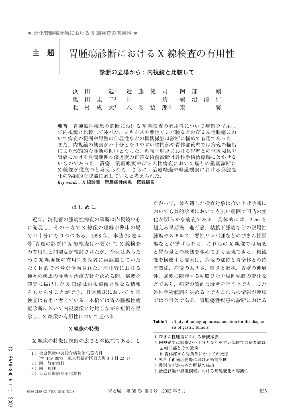
要旨 胃腫瘍性疾患の診断におけるX線検査の有用性について症例を呈示して内視鏡と比較して述べた.スキルスや悪性リンパ腫などのびまん性腫瘍において病変の範囲や胃壁の伸展性などの概観撮影は診断に極めて有用であった.また,内視鏡の観察が不十分となりやすい噴門部や胃体部後壁では病変の描出により形態的な診断の助けとなった.粘膜下腫瘍における胃壁との位置関係や胃癌における浸潤範囲や深達度の正確な術前診断は外科手術治療時に欠かせないものであった.潰瘍,潰瘍瘢痕やびらん性病変において癌との鑑別診断にX線像が役立つと考えられた.さらに,治療経過や経過観察における形態変化の客観的な認識に適していると考えられた.
To assess the utility of radiographic pictures for the diagnosis of gastric tumor, we emphasized its merits and showed cases, comparing X-ray figures with endoscopy. First of all, the radiographic view was useful for gauging the elasticity of the gastric wall and the range of lesions such as scirrhous type of gastric cancer and diffuse type of malignant lymphoma. It was also helpful in detecting tumors located at the cardia and the posterior wall of the gastric body and in showing the width of the lesion and how deeply the cancer had infiltrated the gastric wall. For these reasons, it was necessary to get the X-ray figures before surgical operations for gastric tumors. Another merit of radiography was that it sometimes made it possible to differentiate a carcinoma from a benign erosion, or an ulcer from an ulcer scar because the X-ray figures were able to show mucosal detai ls not obtained by endoscopy. In addition, X-ray pictures were suitable for the objective observation of macroscopic changes evident in follow-up study.

Copyright © 2003, Igaku-Shoin Ltd. All rights reserved.


