Japanese
English
- 有料閲覧
- Abstract 文献概要
- 1ページ目 Look Inside
- 参考文献 Reference
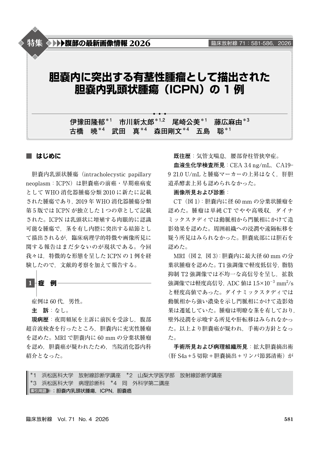
胆嚢内乳頭状腫瘍(intracholecystic papillary neoplasm:ICPN)は胆嚢癌の前癌・早期癌病変としてWHO消化器腫瘍分類2010に新たに記載された腫瘍であり,2019年WHO消化器腫瘍分類第5版ではICPNが独立した1つの章として記載された。ICPNは乳頭状に増殖する肉眼的に認識可能な腫瘍で,茎を有し内腔に突出する結節として描出されるが,臨床病理学的特徴や画像所見に関する報告はまだ少ないのが現状である。今回我々は,特徴的な形態を呈したICPNの1例を経験したので,文献的考察を加えて報告する。
A male patient in his 60s was referred to our hospital after abdominal ultrasonography incidentally revealed a gallbladder mass. Magnetic resonance imaging demonstrated a lobulated intraluminal mass measuring approximately 60 mm in diameter in the gallbladder body. The lesion showed mildly low signal intensity on T1-weighted images, heterogeneous high signal intensity on fat-suppressed T2-weighted images, high signal intensity on diffusion-weighted images, and mildly high signal intensity on the apparent diffusion coefficient map(1.5×10⁻³ mm2/s). Dynamic contrast-enhanced imaging revealed strong arterial enhancement with persistent enhancement into the portal venous phase. A distinct pedunculated stalk was clearly identified, with no evidence of extramural invasion or metastasis. Extended cholecystectomy was performed under suspicion of gallbladder carcinoma, and histopathological examination confirmed intracholecystic papillary neoplasm. This case suggests that careful evaluation of tumor size, the gallbladder wall at the tumor base, mucosal integrity, the presence of a stalk, and the ADC value is important when assessing intraluminal gallbladder masses.
Copyright © 2026, KANEHARA SHUPPAN Co.LTD. All rights reserved.