Japanese
English
- 有料閲覧
- Abstract 文献概要
- 1ページ目 Look Inside
- 参考文献 Reference
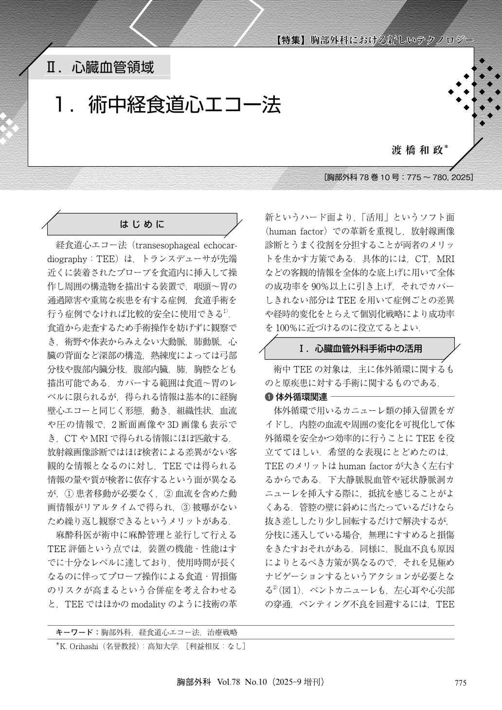
経食道心エコー法(transesophageal echocardiography:TEE)は,トランスデューサが先端近くに装着されたプローブを食道内に挿入して操作し周囲の構造物を描出する装置で,咽頭~胃の通過障害や重篤な疾患を有する症例,食道手術を行う症例でなければ比較的安全に使用できる1).食道から走査するため手術操作を妨げずに観察でき,術野や体表からみえない大動脈,肺動脈,心臓の背面など深部の構造,熟練度によっては弓部分枝や腹部内臓分枝,腹部内臓,肺,胸腔なども描出可能である.カバーする範囲は食道~胃のレベルに限られるが,得られる情報は基本的に経胸壁心エコーと同じく形態,動き,組織性状,血流や圧の情報で,2断面画像や3D画像も表示でき,CTやMRIで得られる情報にほぼ匹敵する.放射線画像診断ではほぼ検者による差異がない客観的な情報となるのに対し,TEEでは得られる情報の量や質が検者に依存するという面が異なるが,①患者移動が必要なく,②血流を含めた動画情報がリアルタイムで得られ,③被曝がないため繰り返し観察できるというメリットがある.
Transesophageal echocardiography (TEE) is a valuable diagnostic and intraoperative tool that allows high-resolution, real-time imaging of deep cardiovascular structures without interfering with surgery. It offers dynamic information similar to computed tomography (CT) or magnetic resonance imaging (MRI) but without radiation exposure, making repeated assessments feasible. During cardiovascular surgery, TEE guides cannula placement, monitors myocardial protection, detects complications like air embolism and intraoperative aortic dissection, and facilitates real-time surgical navigation. Its utility extends to postoperative intensive care unit (ICU) care and emergency settings, where it helps diagnose complications when CT is not feasible. In thoracic surgery, TEE aids in assessing tumor invasion into cardiovascular structures. However, TEE’s effectiveness heavily relies on the operator’s skill, unlike the objectivity of radiologic modalities. Thus, fostering collaboration between anesthesiologists and surgeons is essential. As a critical part of perioperative management, TEE proficiency is now a requirement for board certification in cardiovascular anesthesia in Japan. Supporting anesthesiologists in developing TEE skills enhances surgical outcomes and institutional capability.

© Nankodo Co., Ltd., 2025


