Japanese
English
- 有料閲覧
- Abstract 文献概要
- 1ページ目 Look Inside
- 参考文献 Reference
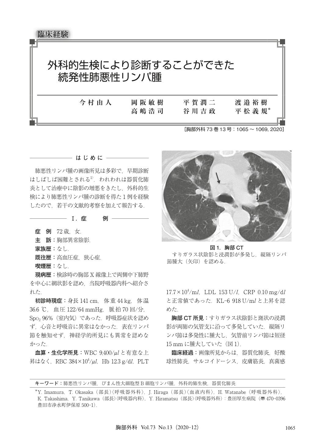
肺悪性リンパ腫の画像所見は多彩で,早期診断はしばしば困難とされる1).われわれは器質化肺炎として治療中に陰影の増悪をきたし,外科的生検により肺悪性リンパ腫の診断を得た1例を経験したので,若干の文献的考察を加えて報告する.
Pulmonary malignant lymphoma presents diverse imaging findings, thus making an imaging-based diagnosis difficult. Furthermore, because of the low histological diagnostic rate of approximately 30% based on transbronchial lung biopsy, there are difficulties in the early diagnosis of pulmonary malignant lymphoma. We report a case of pulmonary malignant lymphoma that was difficult to diagnose until a surgical biopsy was performed. A 72-year-old female was referred to our hospital with an abnormal chest shadow on a medical examination. Chest computed tomography (CT) scan demonstrated ground-glass opacity and consolidation in both lung fields. Bronchoscopy was performed but a histological definitive diagnosis could not be obtained. We suspected organized pneumonia and initiated steroid therapy that resulted in improvement in the chest shadow. However, new multiple lung nodules and mediastinal lymphadenopathy were noticed on CT scan performed 9 months after the initiation of steroid therapy, and a lung biopsy and mediastinal lymph node biopsy were performed. Finally, the diagnosis was malignant lymphoma with pulmonary infiltrates.

© Nankodo Co., Ltd., 2020


