Japanese
English
- 販売していません
- Abstract 文献概要
- 1ページ目 Look Inside
- 参考文献 Reference
- サイト内被引用 Cited by
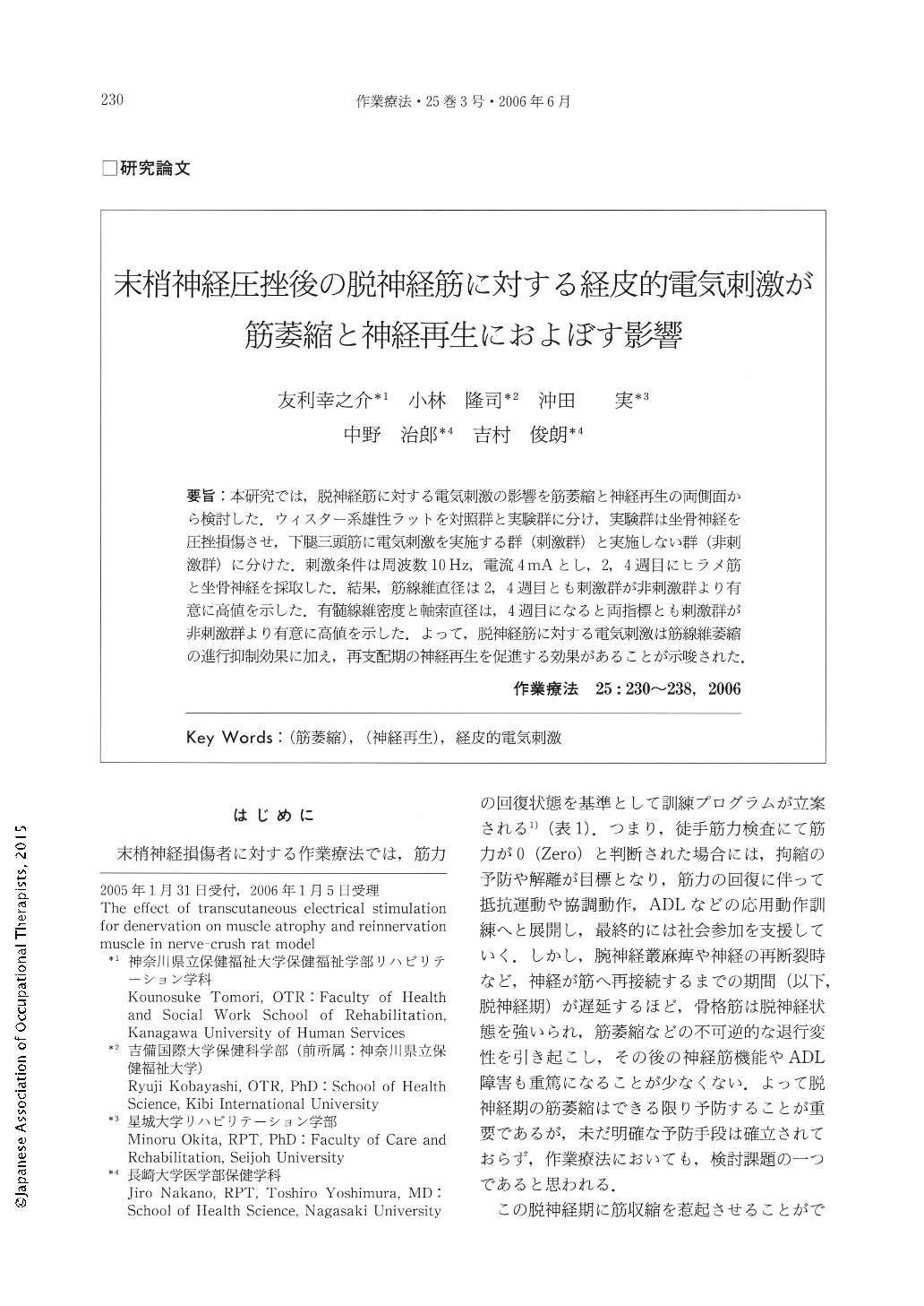
要旨:本研究では,脱神経筋に対する電気刺激の影響を筋萎縮と神経再生の両側面から検討した.ウィスター系雄性ラットを対照群と実験群に分け,実験群は坐骨神経を圧挫損傷させ,下腿三頭筋に電気刺激を実施する群(刺激群)と実施しない群(非刺激群)に分けた.刺激条件は周波数10Hz,電流4mAとし,2,4週目にヒラメ筋と坐骨神経を採取した.結果,筋線維直径は2,4週目とも刺激群が非刺激群より有意に高値を示した.有髄線維密度と軸索直径は,4週目になると両指標とも刺激群が非刺激群より有意に高値を示した.よって,脱神経筋に対する電気刺激は筋線維萎縮の進行抑制効果に加え,再支配期の神経再生を促進する効果があることが示唆された.
The purpose of this study was to determine whether transcutaneous electrical stimulation for denervation muscle effect both muscle atrophy and reinnervation. Eighteen seven-week-old male Wistar rats were divided randomly as follows : control (n = 6), stimulus group (ES group ; n=6), and non-stimulus group (N-ES group ; n=6). In the ES and N-ES groups, a bilateral sciatic nerve was crushed. The triceps surae muscles of rats in the ES group were electrically stimulated, 30 minutes/day for 2, 4 weeks. As a condition of electrical stimulation, the strength was set at 4 Hz, frequencies were set at 10 Hz, and duration was set at 250μsec. Following the experiment, both bilateral soleus muscle and sciatic nerve were removed for histological examination. Mean muscle fiber diameter in the ES group was significantly larger than that of group N-ES both at 2 and 4 weeks. The density of myelinated fibers and mean axon diameter showed no significant difference between the ES group and the N-ES group at 2 weeks, but at 4 weeks, those in the ES group were significantly larger than that of group N-ES. These results suggested that electrical stimulation for denervation muscle inhibits the progress of denervation muscle and leads to regeneration of injury nerve.

Copyright © 2006, Japanese Association of Occupational Therapists. All rights reserved.


