Japanese
English
- 有料閲覧
- Abstract 文献概要
- 1ページ目 Look Inside
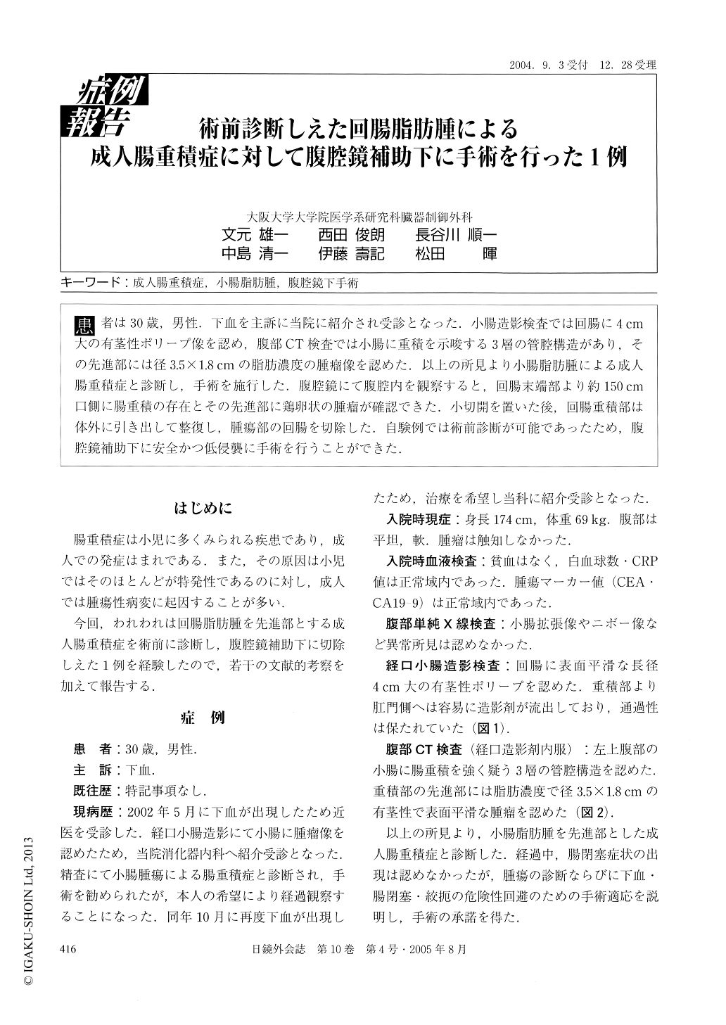
患者は30歳,男性.下血を主訴に当院に紹介され受診となった.小腸造影検査では回腸に4cm大の有茎性ポリープ像を認め,腹部CT検査では小腸に重積を示唆する3層の管腔構造があり,その先進部には径3.5×1.8cmの脂肪濃度の腫瘤像を認めた.以上の所見より小腸脂肪腫による成人腸重積症と診断し,手術を施行した.腹腔鏡にて腹腔内を観察すると,回腸末端部より約150cm口側に腸重積の存在とその先進部に鶏卵状の腫瘤が確認できた.小切開を置いた後,回腸重積部は体外に引き出して整復し,腫瘍部の回腸を切除した.自験例では術前診断が可能であったため,腹腔鏡補助下に安全かつ低侵襲に手術を行うことができた.
A 30-year-old man was admitted to our hospital with melena. Small bowel contrast study showed a 4 cm pedunculated polyp of the ileum. Abdominal computed tomography demonstrated three lumen suggestive of small bowel intussusception and a 3.5×1.8 cm fat density tumor at the leading point. He was, therefore, diag-nosed as adult intussusception due to ileal lipoma and was treated by laparoscopic-assisted surgery. We found the invaginated ileum about 150 cm proximal from the ileum end and an egg-siged tumor at the leading point.

Copyright © 2005, JAPAN SOCIETY FOR ENDOSCOPIC SURGERY All rights reserved.


