Japanese
English
- 有料閲覧
- Abstract 文献概要
- 1ページ目 Look Inside
- 参考文献 Reference
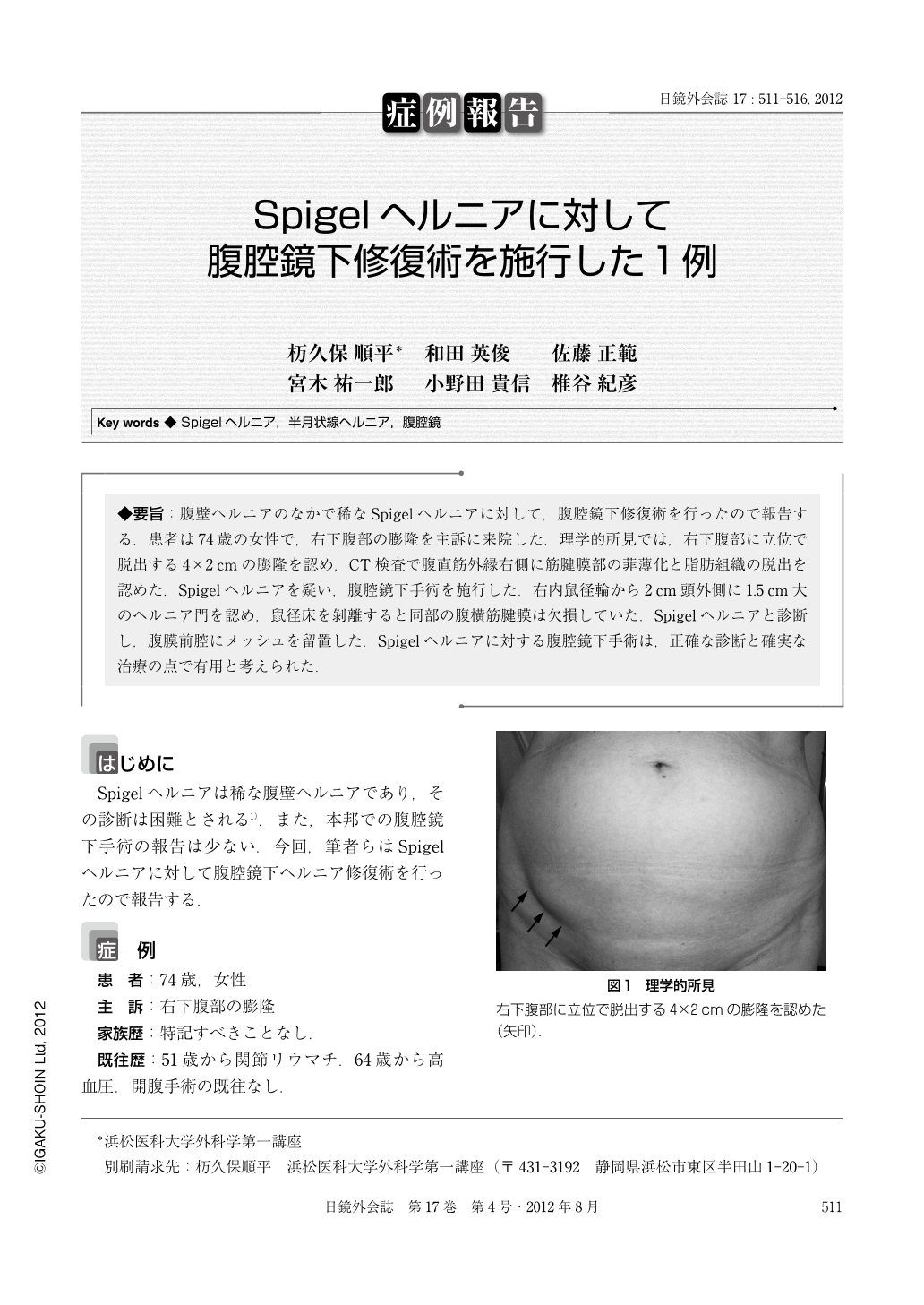
◆要旨:腹壁ヘルニアのなかで稀なSpigelヘルニアに対して,腹腔鏡下修復術を行ったので報告する.患者は74歳の女性で,右下腹部の膨隆を主訴に来院した.理学的所見では,右下腹部に立位で脱出する4×2cmの膨隆を認め,CT検査で腹直筋外縁右側に筋腱膜部の菲薄化と脂肪組織の脱出を認めた.Spigelヘルニアを疑い,腹腔鏡下手術を施行した.右内鼠径輪から2cm頭外側に1.5cm大のヘルニア門を認め,鼠径床を剝離すると同部の腹横筋腱膜は欠損していた.Spigelヘルニアと診断し,腹膜前腔にメッシュを留置した.Spigelヘルニアに対する腹腔鏡下手術は,正確な診断と確実な治療の点で有用と考えられた.
Spigelian hernia is a rare abdominal wall hernia which is generally difficult to diagnose. There are only few reports of laparoscopic Spigelian hernia repair in Japan. A 74-year-old woman visited our hospital because of a bulge on the right lower quadrant. There was a 4×2cm bulge at standing position. Computed tomography showed a thinning of musculoaponeurotic layers at the right outer side of rectus abdominis muscle and a protrusion of fat tissue. Spigelian hernia was suspected and laparoscopic repair was performed. Laparoscopically, the hernia orifice was 1.5×0.5cm at cranial and lateral site, 2cm away from the right internal inguinal ring. The inguinal floor was dissected, and the defect of transverses abdominis aponeurosis was found, confirming the diagnosis of Spigelian hernia. A 13×9cm polyester mesh was placed into the preperitoneal space with tacker fixation. The peritoneum was closed with interrupted sutures. Laparoscopic surgery is useful for the definite diagnosis and the repair of Spigelian hernia, especially for those that are difficult to diagnose by physical examination and imaging findings.

Copyright © 2012, JAPAN SOCIETY FOR ENDOSCOPIC SURGERY All rights reserved.


