Japanese
English
- 有料閲覧
- Abstract 文献概要
- 1ページ目 Look Inside
- 参考文献 Reference
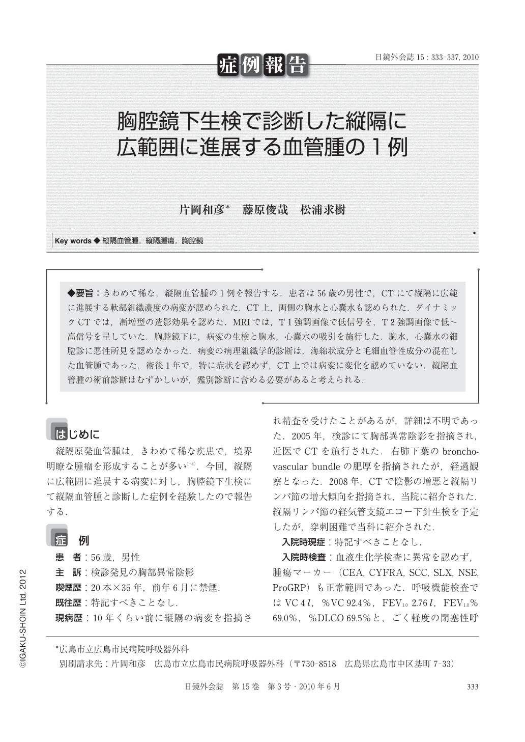
◆要旨:きわめて稀な,縦隔血管腫の1例を報告する.患者は56歳の男性で,CTにて縦隔に広範に進展する軟部組織濃度の病変が認められた.CT上,両側の胸水と心囊水も認められた.ダイナミックCTでは,漸増型の造影効果を認めた.MRIでは,T1強調画像で低信号を,T2強調画像で低~高信号を呈していた.胸腔鏡下に,病変の生検と胸水,心囊水の吸引を施行した.胸水,心囊水の細胞診に悪性所見を認めなかった.病変の病理組織学的診断は,海綿状成分と毛細血管性成分の混在した血管腫であった.術後1年で,特に症状を認めず,CT上では病変に変化を認めていない.縦隔血管腫の術前診断はむずかしいが,鑑別診断に含める必要があると考えられる.
A rare case of mediastinal hemangioma was reported in this article. A 56-year-old man was found to have a lesion of soft tissue density widely infiltrating to the mediastinum on CT scans. CT also showed bilateral pleural and pericardial effusion. On contrast-enhanced dynamic CT, gradually increasing enhancement was found in the lesion. The lesion showed low intensity on T 1-weighted magnetic resonance imaging and low to high intensity on T 2. Biopsy of the lesion and aspiration of the pleural and pericardial effusion were performed under the video-assisted thoracic surgery. Cytologic examination of the pleural and pericardial effusion showed no malignancy. The histopathological diagnosis of the lesion was cavernous and papillary hemangioma. The patient had no symptoms and the lesion had no changes on the CT scans for a year. Althogh a preoperative diagnosis of mediastinal hemangioma is difficult, such tumors should be included in the differential diagnosis.

Copyright © 2010, JAPAN SOCIETY FOR ENDOSCOPIC SURGERY All rights reserved.


