Japanese
English
- 有料閲覧
- Abstract 文献概要
- 1ページ目 Look Inside
- 参考文献 Reference
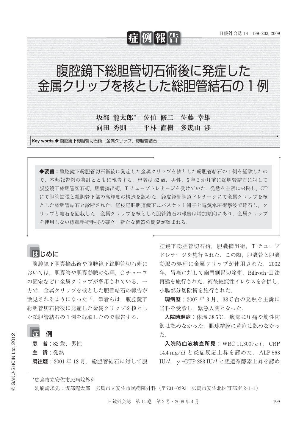
◆要旨:腹腔鏡下総胆管切石術後に発症した金属クリップを核とした総胆管結石の1例を経験したので,本邦報告例の集計とともに報告する.患者は82歳,男性.5年3か月前に総胆管結石に対して腹腔鏡下総胆管切石術,胆囊摘出術,Tチューブドレナージを受けていた.発熱を主訴に来院し,CTにて胆管拡張と総胆管下部の高輝度の構造を認めた.経皮経肝胆道ドレナージにて金属クリップを核とした総胆管結石と診断された.経皮経肝胆道鏡下にバスケット鉗子と電気水圧衝撃波で砕石し,クリップと結石を回収した.金属クリップを核とした胆管結石の報告は増加傾向にあり,金属クリップを使用しない標準手術手技の確立,新たな機器の開発が望まれる.
We report a case of common bile duct stone containing a metallic clip after laparoscopic common bile duct exploration and review the Japanese literature. An 82-year-old man was urgently admitted to our hospital for high fever. He previously underwent laparoscopic common bile duct exploration, cholecystectomy, and T tube drainage for common bile duct stone in our hospital 5 years and 3 months ago. Abdominal computed tomography showed dilatation of the bile duct and a high density structure at the inferior part of the common bile duct. Percutaneous transhepatic biliary drainage was performed and cholangiography showed a common bile duct stone containing a metallic clip. The stone and metallic clip was removed using a basket catheter and electrohydraulic shock wave lithotomy under percutaneous transhepatic cholangioscopy. Case reports of common bile duct stone due to metallic clip migration has increased recently. Establishment of a standard operative procedure of laparoscopic cholecystectomy without using metallic clips and development of new instrument are expected.

Copyright © 2009, JAPAN SOCIETY FOR ENDOSCOPIC SURGERY All rights reserved.


