Japanese
English
- 有料閲覧
- Abstract 文献概要
- 1ページ目 Look Inside
I.はじめに
放線菌症は慢性に経過する化膿性肉芽腫性疾患で,1845年Langcnbeckが脊椎カリエスの患者の膿汁より本症に特有な菌塊を発見したのが最初とされている。比較的稀な疾患であり,さらに近年における抗生物質の発達と普及に伴い日常臨床で本症に遭遇することが少なくなった。その中で頸部・顔面領域は好発部位の1つとされるが,そのほとんどは歯科領域からの感染により下顎部,頸部に発症するものである。
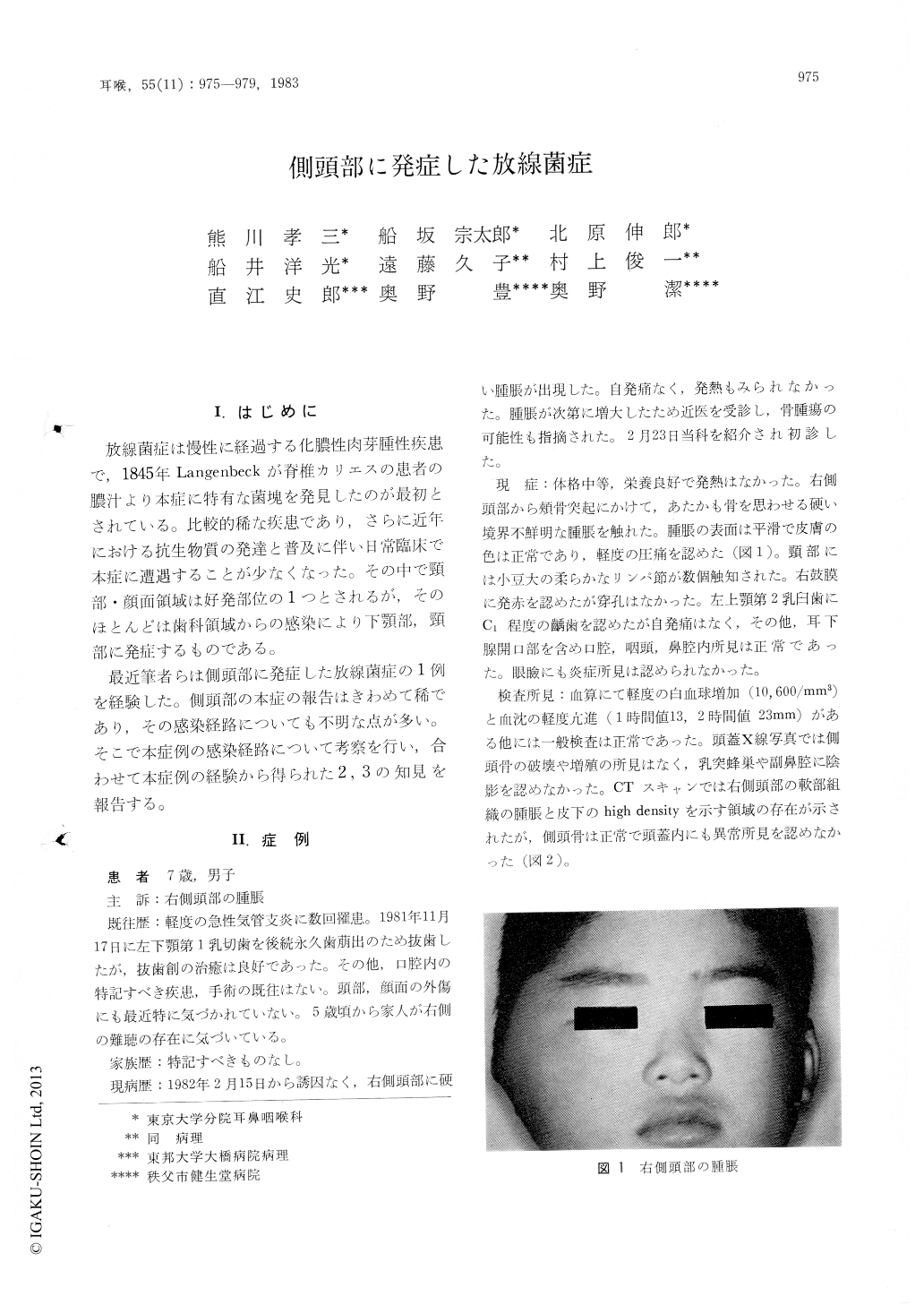
最近筆者らは側頭部に発症した放線菌症の1例を経験した。側頭部の本症の報告はきわめて稀であり,その感染経路についても不明な点が多い。そこで本症例の感染経路について考察を行い,合わせて本症例の経験から得られた2,3の知見を報告する。
The patient is a 7-year-old boy with the complaint of swelling of the right temporal region. An exploratory incision revealed that a large amount of yellowish pus containing the characteristic sulfar granules was found in the abscess cavity located in the subcutaneous space. Microscopic observation of the sulfar granule showed figures of actinomycotic infection. A pertinent dosis of penicillin was administrated for 15 weeks with good results.
Actinomycosis of the temporal region is very rare, because the most common cause of the cervicofacial actinomycosis is dental traction and teeth caries. The possible routes for contamination of actinomyces were discussed in this case. Since no definite promoting episode of trauma was recognized, the middle ear seemed one of the most probable portals of entry in consequence of prior recognition of acute otitis media.

Copyright © 1983, Igaku-Shoin Ltd. All rights reserved.


