Japanese
English
- 有料閲覧
- Abstract 文献概要
- 1ページ目 Look Inside
I.はじめに
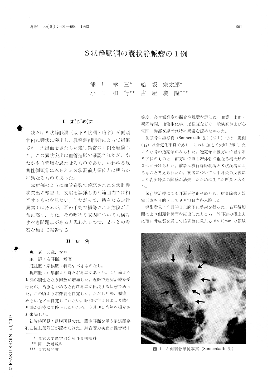
我々はS状静脈洞(以下S状洞と略す)が側頭骨内に嚢状に突出し,乳突洞削開術によって損傷され,大出血をきたした走行異常の1例を経験した。この嚢状突出は血管造影で確認されたが,あたかも血管瘤を思わせるものであり,いわゆる危側性側頭骨にみられるS状洞前方偏位とは明らかに異なるものであった。
本症例のように血管造影で確認されたS状洞嚢状突出の報告は,文献を渉猟し得た範囲内では相当するものを見ない。したがって,稀有なる走行異常ではあるが,耳の手術で損傷される危険が非常に高く,また,その呼称や成因についても検討すべき問題点があると思われるので,2〜3の考察を加えて報告する。
A rare case of a saccular venous aneurysm of the sigmoid sinus (SS) is reported.
The patient is a 57-year-old female who was diagnosed as having chronic otitis media of the right ear. On plain x-ray films of the temporal bone, a lucent area was noted in the mastoid area, suggesting a very large cell overlying the knee of SS. Tympanoplasty was performed, and then the abnormal enlargement of SS was found during the exploratory mastoidectomy.
Serial carotid angiograms showed no abnormality on the arterial phase but revealed a saccular protrusion of SS into the temporal bone on the venous phase. This protrusion almost occupied the whole mastoid cavity and expanded to the cortical bone. However, neither bruit nor thrill was recognized in the retroauricular region on careful examination.
The authors named this disease as "saccular venous aneurysm of SS". The otologic surgeon should remind of this disease on seeing an abnormal lucent area in the mastoid area and "blue mastoid cortex" in order to avoid the injury of SS during mastoidectomy.

Copyright © 1983, Igaku-Shoin Ltd. All rights reserved.


