Japanese
English
- 有料閲覧
PPV購入案内
1,320円 (1,200円+税10%) こちらは、97ページから102ページの文献です。購入には医書.jp本会員登録が必要です。
会員登録完了後に改めて上記「購入サイトへ」を選択してください。
または「購入サイトへ」選択後に「購入ログイン」、「新規会員登録」の順に進んでください。
会員登録について詳しくはをご確認ください。
- Abstract 文献概要
- 1ページ目 Look Inside
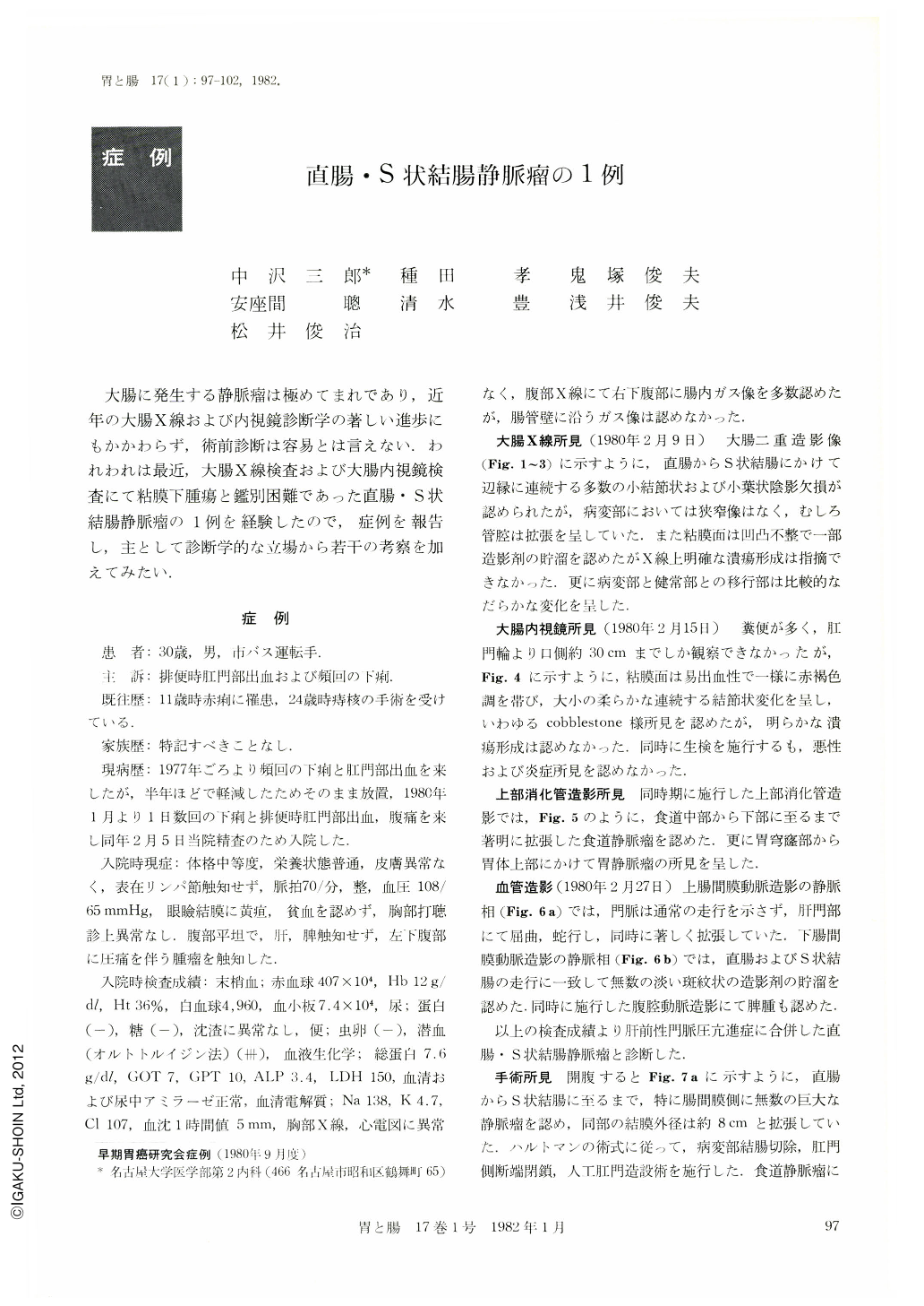
大腸に発生する静脈瘤は極めてまれであり,近年の大腸X線および内視鏡診断学の著しい進歩にもかかわらず,術前診断は容易とは言えない.われわれは最近,大腸X線検査および大腸内視鏡検査にて粘膜下腫瘍と鑑別困難であった直腸・S状結腸静脈瘤の1例を経験したので,症例を報告し,主として診断学的な立場から若干の考察を加えてみたい.
The patient was a 30-year-old man with chief complaints of diarrhea, lower abdominal pain and bloody stool since two or three years ago. The patient admitted to our hospital, because these symptoms were aggravated. Barium enema examination was grossly abnormal showing marginal irregularity on the rectum and sigmoid colon. Double contrast picture showed cobblestone-like appearance.
Colonoscopic findings showed multiple soft nodular elevation and no ulceration on the rectosigmoid colon. Abdominal angiography revealed the pooling of the contrast medium along the rectosigmoid colon and dilated tortous portal vein in the venous phase.
The patient was diagnosed as prehepatic portal hypertension and colonic varices. UGI series showed varices on the esophagus and upper area of the stomach. Hartmann's method operation and proximal gastric devascularization were performed. Varices of the colon are very rare and there have been 31 additional cases in world literature.
Copyright © 1982, Igaku-Shoin Ltd. All rights reserved.