Japanese
English
- 有料閲覧
- Abstract 文献概要
- 1ページ目 Look Inside
I.はじめに
Yoshieら1)やAranらなどにより始められた蝸電図も現在では広く臨床検査として用いられており,内耳疾患の診断の前要な検査のり一つとなっている。この蝸電図所見から内耳病態の解明や疾患の細別診断を日的とする試みは多数報告されているが,いまだその目的を達したとはいえない。蝸電図は内耳に関する多数の情報を知らせてくれるが,そのうちの一つにAPのN1潜時の問題があるものと思われる。他の情報(例えば振幅の入出力曲線,波形,SP)に比べて潜時ば記録条件に左右されることなく常に一定の結果が得られるので注目すべき現象と思われる。
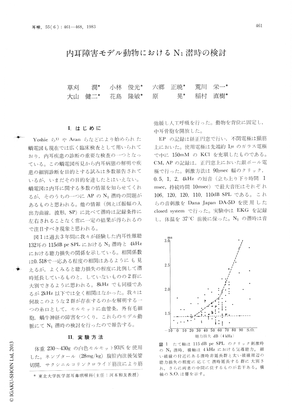
図1は過去3年間に我々が経験した内耳性難聴132耳の115dB pe SPLにおけるN1潜時と4kHzにおける聴力損失の関係を示している。相関係数は0.518で一応ある程度の相関はあるようにも見えるが,よくみると聴力損失の程度に比例して潜時延長しているものと,していないものの2群に大別できるように思われる。8kHzでも同様であるが2kHz以下では全く相関はなかった。我々は何故このような2群が存在するのかを解明する一つの糸口として,モルモットに血管条,外有毛細胞,蝸牛神経の障害をつくり,これらのモデル動脈にてN1潜時の検討を行ったので報告する。
It is well known that some sensorineural hearing loss exhibits N1latency prolongation but some does not. To clarify this phenomenon, the N1latency was examined in the guinea pigs subjected to temporal anoxia or given ethacrynic acid, kanamycin and kainic acid, and the following results were obtained.
1) Ethacrynic acid and kainic acid did not produce the N1latency prolongation.
2) The N1latency was prolonged in the severely damaged animals by kanamycin and temporal anoxia.
The results obtained in the present study suggest that the N1latency is prolonged in cases of sensory hair cell damage and not prolonged in the dysfunction of the stria vascularis or the cochlear nerve.

Copyright © 1983, Igaku-Shoin Ltd. All rights reserved.


