Japanese
English
- 有料閲覧
- Abstract 文献概要
- 1ページ目 Look Inside
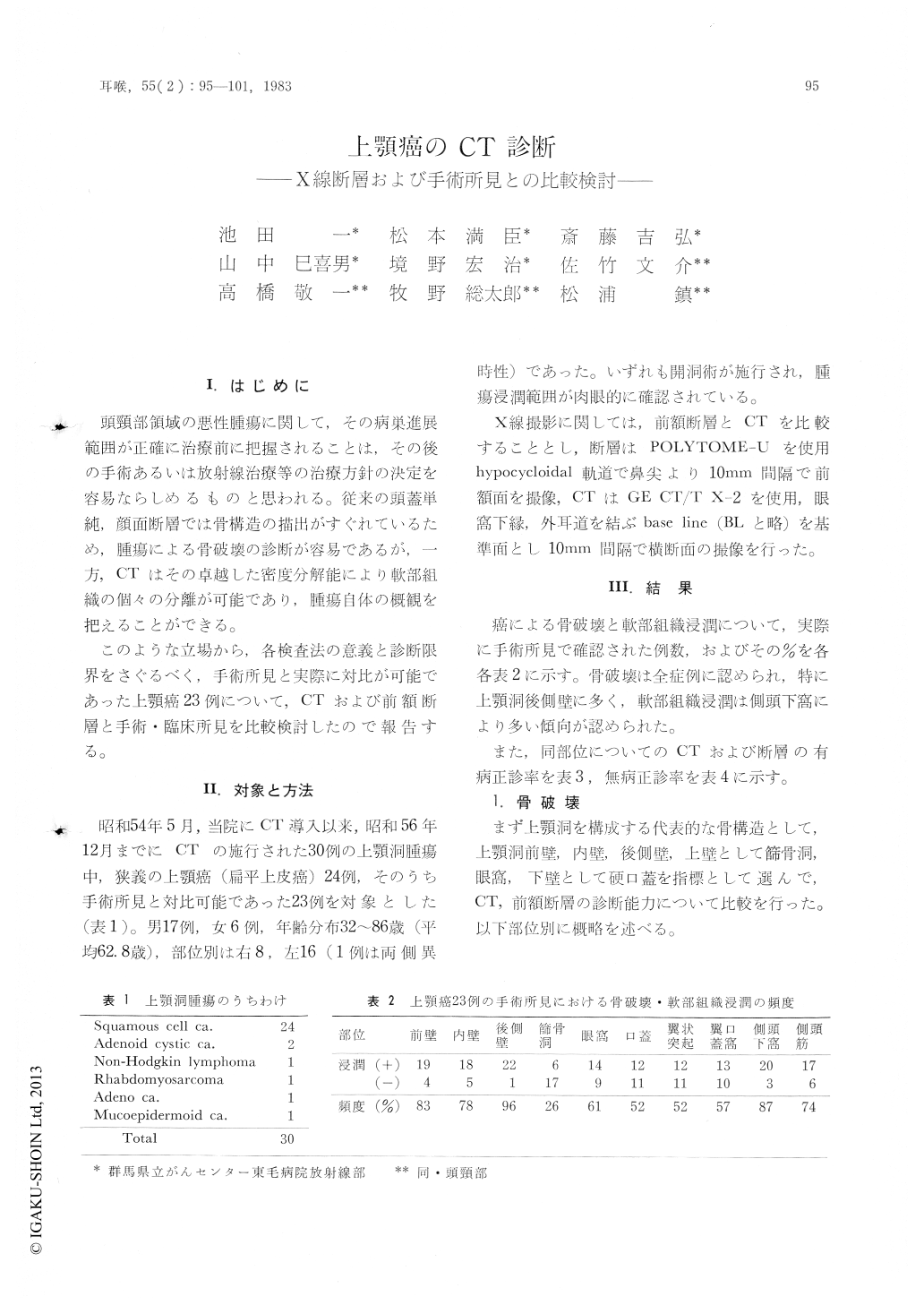
I.はじめに
頭頸部領域の悪性腫瘍に関して,その病巣進展範囲が正確に治療前に把握されることは,その後の手術あるいは放射線治療等の治療方針の決定を容易ならしめるものと思われる。従来の頭蓋単純,顔面断層では骨構造の描出がすぐれているため,腫瘍による骨破壊の診断が容易であるが,一方,CTはその卓越した密度分解能により軟部組織の個々の分離が可能であり,腫瘍自体の概観を把えることができる。
このような立場から,各検査法の意義と診断限界をさぐるべく,手術所見と実際に対比が可能であった上顎癌23例について,CTおよび前額断層と手術・臨床所見を比較検討したので報告する。
Twenty three patients of maxillary cancer were examined by computed tomography and conventional frontal tomography, and their diagnostic accuracy was investigated.
Accuracy of computed tomography for detection of bone destruction is almost equal to that of tomography, through superior to tomography in recognition of tumor extent, at both pterygopalatine fossa and infratemporal fossa.
We must be careful in evaluation of invasion to the ethmoid sinus, because of many false positives.
The computed tomographic images are useful for management of maxillary cancer, not only in treatment planning but also in follow up examination after combination therapy.

Copyright © 1983, Igaku-Shoin Ltd. All rights reserved.


