Japanese
English
- 有料閲覧
- Abstract 文献概要
- 1ページ目 Look Inside
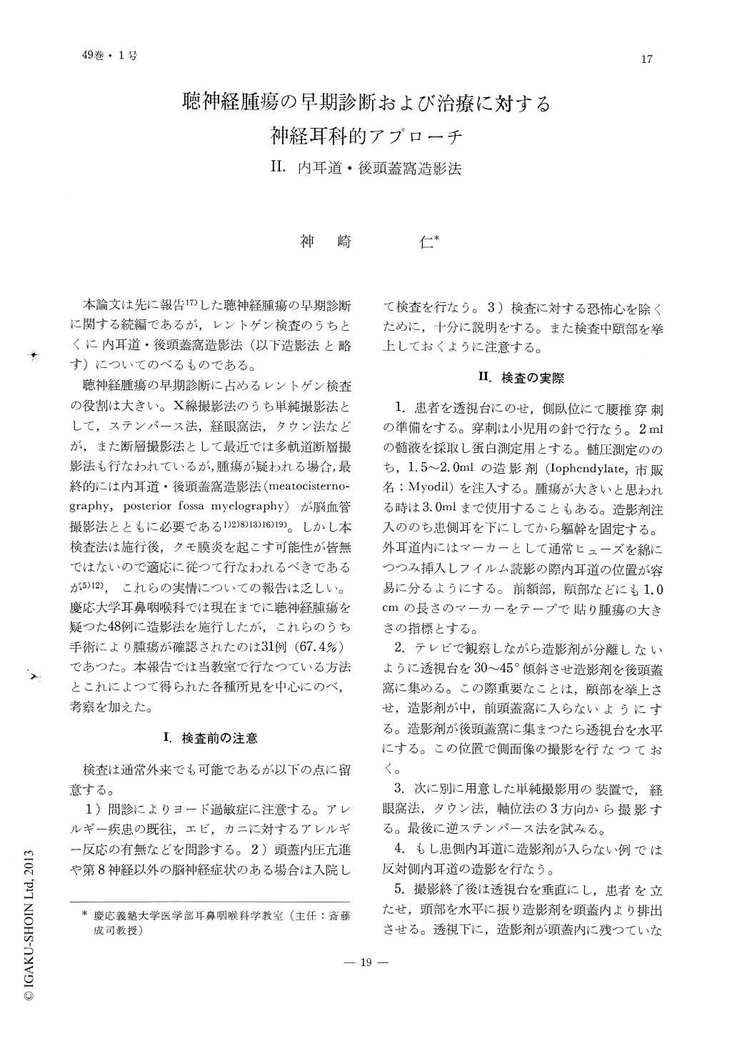
本論文は先に報告17)した聴神経腫瘍の早期診断に関する続編であるが,レントゲン検査のうちとくに内耳道・後頭蓋窩造影法(以下造影法と略す)についてのべるものである。
聴神経腫瘍の早期診断に占めるレントゲン検査の役割は大きい。X線撮影法のうち単純撮影法として,ステンバース法,経眼窩法,タウン法などが,また断層撮影法として最近では多軌道断層撮影法も行なわれているが,腫瘍が疑われる場合,最終的には内耳道・後頭蓋窩造影法(meatocisternography,posterior fossa myelography)が脳血管撮影法とともに必要である1)2)8)13)16)19)。しかし本検査法は施行後,クモ膜炎を起こす可能性が皆無ではないので適応に従つて行なわれるべきであるが5)12),これらの実情についての報告は乏しい。慶応大学耳鼻咽喉科では現在までに聴神経腫瘍を疑つた48例に造影法を施行したが,これらのうち手術により腫瘍が確認されたのは31例(67.4%)であつた。本報告では当教室で行なつている方法とこれによつて得られた各種所見を中心にのべ,考察を加えた。
Our method of technique in differentiating the normal and pathological picture by meatocisternography was presented. This method should be applied in accordance to the conditions of the case and at present Sheehy's rule appears to be the best to be followed. In cases of Rechlinghausen disease devoid of cerebral nerve symptoms and without eighth nerve involvement, this method of exposure may often reveal a latent tumor.
Of the 48 cases of myelographies performed in this way, which were followed by operative procedures, there were 31 cases (67.4%) of confirmed acoustic tumors.
False positive cases are also taken into discussion.

Copyright © 1977, Igaku-Shoin Ltd. All rights reserved.


