Japanese
English
- 有料閲覧
- Abstract 文献概要
- 1ページ目 Look Inside
I.はじめに
無症候性の傍床突起内頸動脈動脈瘤の増加につれて,顕微鏡手術例も増加している12,14,17).この部位の動脈瘤は変化に富んだ解剖構築に囲まれているため,他の部位のそれと比較して手術は一般に困難である.しかし解剖学的変化とそれに由来する手術の困難度,予想される合併症と対処法などについては既に多くの報告があり2,5,7,9,10,21-23,26),術前にこれらの点を検討することにより顕微鏡手術の成績を向上させることができると考えられる.
一方,血管内手術による動脈瘤内コイル塞栓術(以下血管内手術と略す)は近年著しく普及してきた.この部位の動脈瘤は彎曲部に存在し,また術中の塞栓操作のみならず手術終了後もサイホン部内頸動脈の複雑で大きな血流の影響を受ける.このため他の部位とは別の困難要素があり20,25),またより長期的な追跡が不可欠とされている.しかし血管内手術は血管外の頭蓋底構築の変化に影響されにくいため,この部位の動脈瘤の治療に適しているとも考えられ,最近その治療例が増加している6,20,24,25).
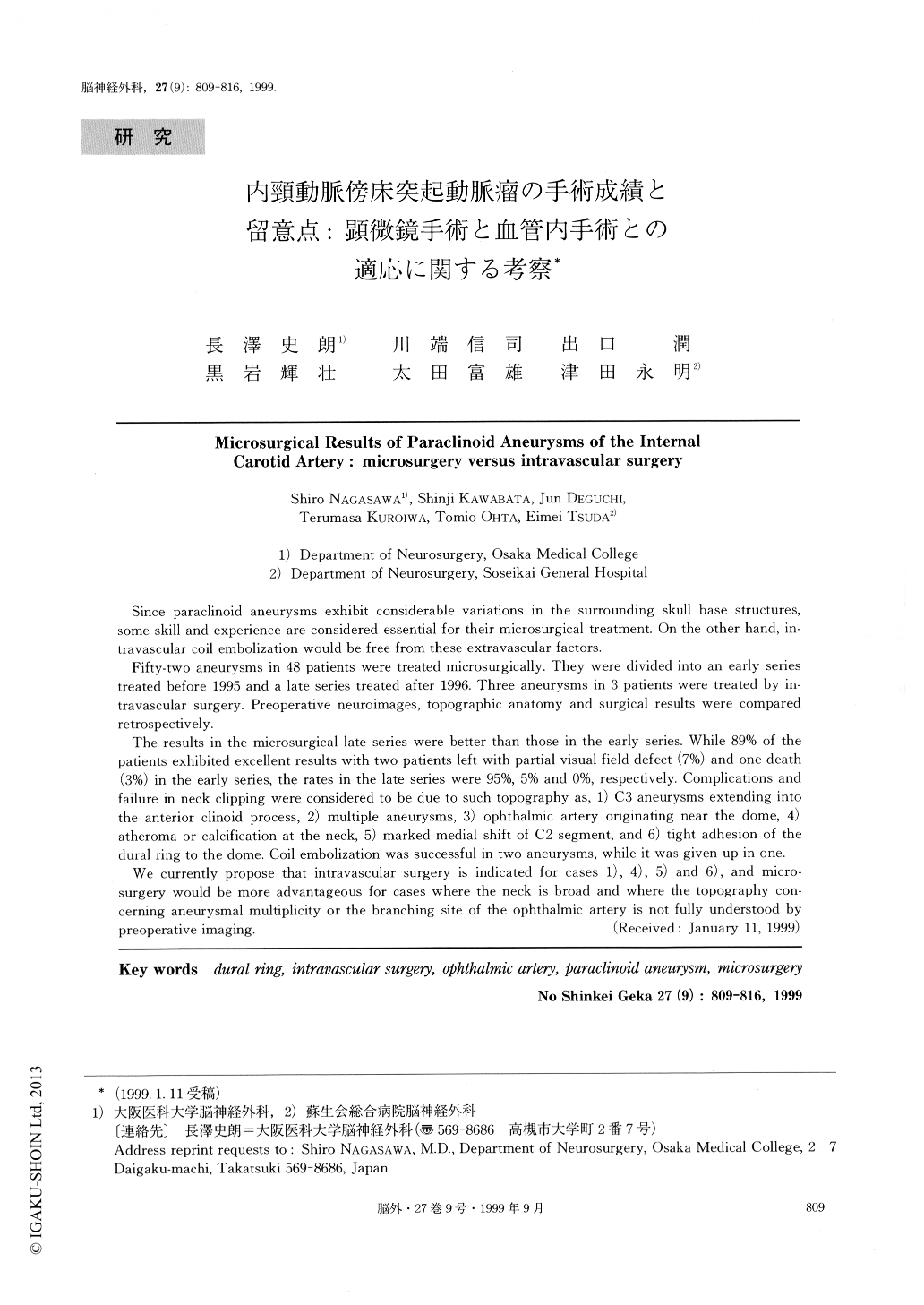
Since paraclinoid aneurysms exhibit considerable variations in the surrounding skull base structures,some skill and experience are considered essential for their microsurgical treatment. On the other hand, in-travascular coil embolization would be free from these extravascular factors.
Fifty-two aneurysms in 48 patients were treated microsurgically. They were divided into an early seriestreated before 1995 and a late series treated after 1996. Three aneurysms in 3 patients were treated by in-travascular surgery. Preoperative neuroimages, topographic anatomy and surgical results were comparedretrospectively.
The results in the microsurgical late series were better than those in the early series. While 89% of thepatients exhibited excellent results with two patients left with partial visual field defect (7%) and one death(3%) in the early series, the rates in the late series were 95%, 5% and 0%, respectively. Complications andfailure in neck clipping were considered to be due to such topography as, 1) C3 aneurysms extending intothe anterior clinoid process, 2) multiple aneurysms, 3) ophthalmic artery originating near the dome, 4)atheroma or calcification at the neck, 5) marked medial shift of C2 segment, and 6) tight adhesion of thedural ring to the dome. Coil embolization was successful in two aneurysms, while it was given up in one. We currently propose that intravascular surgery is indicated for cases 1), 4), 5) and 6), and micro-surgery would be more advantageous for cases where the neck is broad and where the topography con-cerning aneurysmal multiplicity or the branching site of the ophthalmic artery is not fully understood bypreoperative imaging.

Copyright © 1999, Igaku-Shoin Ltd. All rights reserved.


