Japanese
English
- 有料閲覧
- Abstract 文献概要
- 1ページ目 Look Inside
I.はじめに
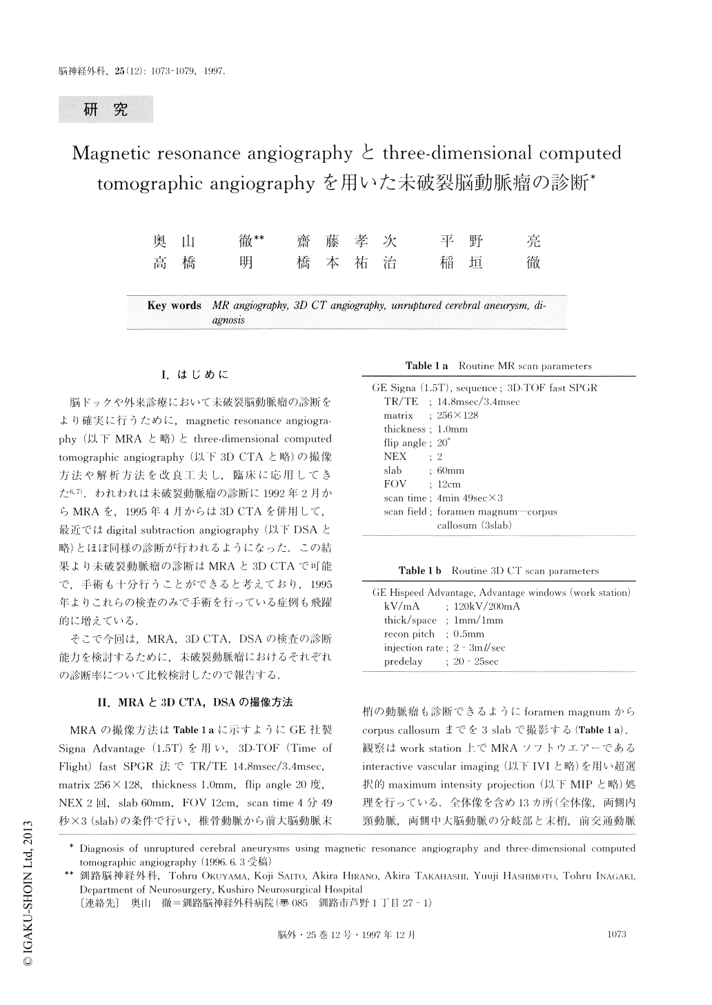
脳ドックや外来診療において未破裂脳動脈瘤の診断をより確実に行うために,magnetic resonance angiogra—phy(以下MRAと略)とthree-dimensional computed tomographic angiography(以下3D CTAと略)の撮像方法や解析方法を改良工夫し,臨床に応用してきた6,7).われわれは未破裂動脈瘤の診断に1992年2月からMRAを,1995年4月からは3D CTAを併用して,最近ではdigital subtraction angiography(以下DSAと略)とほぼ同様の診断が行われるようになった.この結果より未破裂動脈瘤の診断はMRAと3D CTAで可能で,手術も十分行うことができると考えており,1995年よりこれらの検査のみで手術を行っている症例も飛躍的に増えている.
そこで今回は,MRA,3D CTA,DSAの検査の診断能力を検討するために,未破裂動脈瘤におけるそれぞれの診断率について比較検討したので報告する.
The purpose of this study is to confirm the use of magnetic resonance (MR) angiography and three- dimensional computed tomographic (3D CT) angiogra- phy, in the screening of unruptured cerebral aneurysms.
Sixty-six unruptured cerebral aneurysms in forty-eight patients were examined by MR angiography, 3D CT angiography and digital subtraction angiography (DSA). All cases underwent surgery. Three out of six-ty-six (4.5%) cerebral aneurysms detected by MR angiography and 3D CT angiography were false posi-tive. The false aneurysms were located at the region of the internal carotid artery and the posterior communi-cating artery, and were under 2.0mm in size. The di-agnosis of the infundibular dilatation at the junction of the internal carotid artery and the posterior communi-cating artery remained difficult. The true positive di-agnosis of aneurysms using MR angiography or 3D CT angiography was 95.5%, in our hospital. All unruptured cerebral aneurysms over 2.0mm in size were detected correctly by using MR angiography and 3D CT angiography, as well as DSA. The inadequate diagnosis of aneurysms caused by MR angiography was due to the overlapping of vessels and the surrounding noise. Superselective maximum intensity projection (MIP) and interactive vascular imaging (IVI) were adopted for exact diagnosis. Furthermore, the three slab method of MR angiography was used for containing the limits between the vertebral artery and the distal anterior cerebral artery, and the triple method was able to de-crease surrounding noise. Using MR angiography, we diagnosed and operated on about 100 cases of unrup-tured cerebral aneurysm in one year.
Our conclusion is that we can diagnose any unrup-tured cerebral aneurysm, over 2.0mm in size, using MR angiography and 3D CT angiography.

Copyright © 1997, Igaku-Shoin Ltd. All rights reserved.


