Japanese
English
- 有料閲覧
- Abstract 文献概要
- 1ページ目 Look Inside
I.はじめに
乳児期発症の中枢神経系腫瘍は稀であり,かつ同時期に発生する悪性脳腫瘍は,年長児と比べ治療は難渋し,予後も不良である2,4,7).今回われわれは,乳児期松果体部に発生した脳腫瘍に手術,化学療法を施行し一過性の腫瘍縮小効果を得たが,その後間もなくして,急速な腫瘍再増大を来たした症例を経験した.その臨床経過,病理組織所見から中枢神経系発生のrhabdoid tumorと診断した.同疾患は乳幼児期に発生する極めて悪性経過をたどる腫瘍であり,近年欧米からの報告が相次いでいる5,6,9-11).しかし,本邦からの報告はわれわれが渉猟し得た中で1例のみであるため13),同疾患について若干の文献的考察を加え報告する.
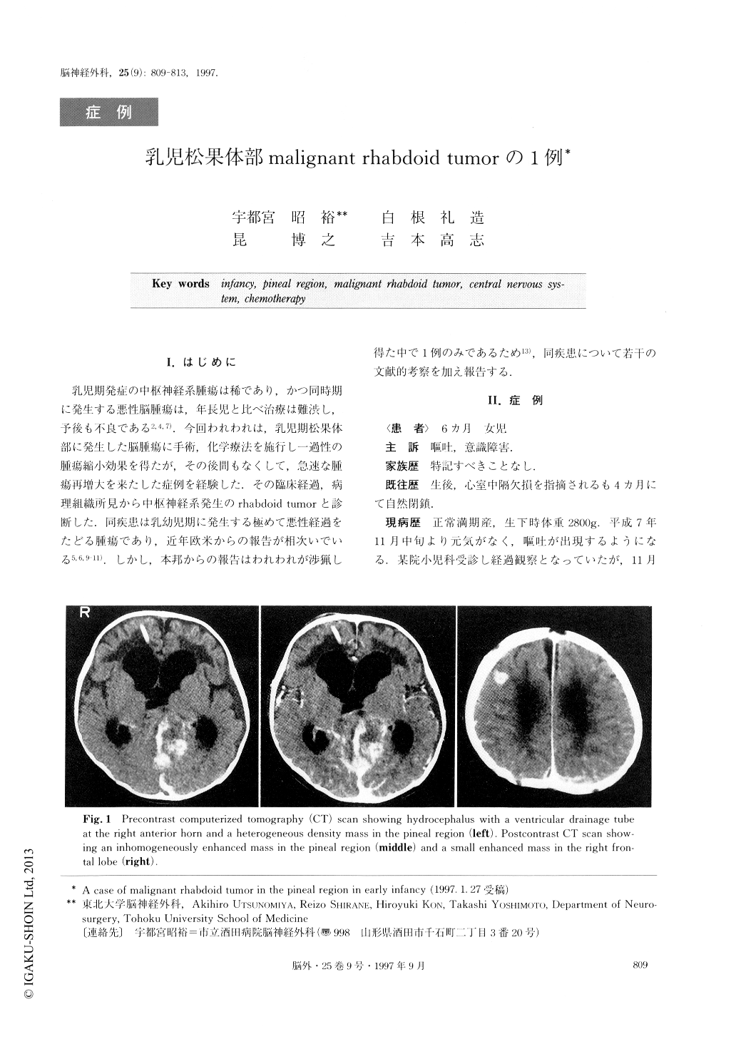
A 6-month-old girl was admitted to another hospital because of consciousness disturbance, preceded by 2 weeks of decreased activity and vomiting. She was re-ferred to our hospital after ventricular drainage had been instituted for hydrocephalus and the tumor in the pineal region. The patient was noted to have conju-gate upward gaze palsy and papilledema. CT scan and MRI revealed a large tumor in the pineal region with tumoral hemorrhage and a small mass in the right fron-tal lobe. At surgery, the pineal region tumor was re-moved subtotally. Histological examination showed the tumor to be composed of sheets of large polyhedral or round cells with an eccentric round nuclei, prominent nucleoli, and cytoplasmic inclusions. Immunohistoche-mical studies were positive for GFAP, vimentin, S-100, CK, EMA, and SMA, but negative for AFP, HCG, PLAP, and CEA.
Following surgery, she received three 5-day cycles of chemotherapy, consisting of intravenous administration of cisplatin 20 mg/m2/day and etoposide 60mg/m2/ day. After these therapies, MRI showed a decrease in the area of high intensity in the pineal region, but almost no change in the right frontal mass lesion. Fol-low-up radiological examination showed that the tumor had grown rapidly one month after chemotherapy and the patient died 5 months after her first hospitalization. Malignant rhabdoid tumor of the CNS is rare and re-markably malignant. This tumor should be treated us-ing multidisciplinary management with surgery, inten-sive chemotherapy, and radiotherapy depending on the patient's age.

Copyright © 1997, Igaku-Shoin Ltd. All rights reserved.


