Japanese
English
- 有料閲覧
- Abstract 文献概要
- 1ページ目 Look Inside
I.はじめに
耳鼻咽喉科領域においてよく知られているコレステリン肉芽腫cholesterol granulomaは,種々の原因で発生したコレステリン結晶に対する防御組織反応により形成される肉芽腫である.本肉芽腫の好発部位は中耳腔であるが3),稀に錐体骨尖端部に発生することがあり1),この場合には脳神経外科の対象疾患となる.われわれは,錐体骨尖端部のcholesterol granulomaを経験したので,臨床像,特に画像診断及び治療に関して,若干の考察を加えて報告する.
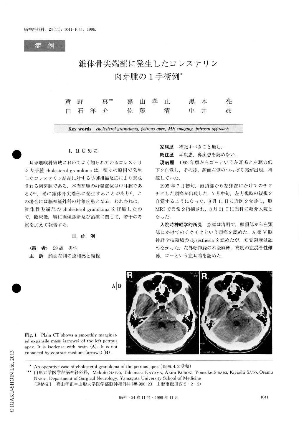
A 59-year-old man presented with a rare cholesterol granuloma of the petrous apex manifesting as headache, left facial dysesthesia, diplopia, left hearing impairment, and left tinnitus. Neurological examination revealed dysesthesia of territory in all divisions of the left trigeminal nerve, left incomplete abducens nerve palsy, left mixed hearing impairment, and left tinnitus. Plain CT scan showed a smoothly marginated mass in-volving the left petrous apex. The mass was isodense with the brain parenchyma and not enhanced by con-trast medium. The mass appeared heterogeneously slightly hyperintense on the T1-weighted MR image and homogeneously hyperintense on the T2-weighted MR image except for the peripheral portion. The mass was not enhanced after intravenous gadolinium DTPA administration. Surgery via a petrosal approach totally removed the mass in the intracranial, extradural space. Histological examination showed typical features of cholesterol granuloma, with cholesterin clefts, hemo-siderin deposits, and erythrocytes in non-specific gra-nulation tissue. Cholesterol granuloma most commonly occurs in the middle ear cavity, and rarely in the pet-rous apex. The characteristic hyperintense appearance of cholesterol granuloma on T1- and T2-weighted MR images is very useful for differentiation from other le-sions of the petrous apex and the cerebellopontine angle such as cholesteatoma, mucocele, chordoma, and meningioma. Solid cholesterol granuloma of the petrous apex should be treated by total removal via craniotomy, not by drainage which is commonly performed by otorhinologists.

Copyright © 1996, Igaku-Shoin Ltd. All rights reserved.


