Japanese
English
- 有料閲覧
- Abstract 文献概要
- 1ページ目 Look Inside
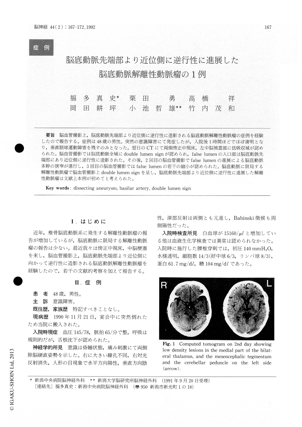
脳血管撮影上,脳底動脈先端部より近位側に逆行性に造影される脳底動脈解離性動脈瘤の症例を経験したので報告する。症例は48歳の男性。突然の意識障害にて発症したが,入院後1時間ほどでほぼ清明となり,垂直眼球運動障害を残すのみとなった。翌日のCTにて両側傍正中視床,左中脳被蓋部に低吸収域が認められた。脳血管撮影では脳底動脈全域にdouble lumen signが認められ,false lumenの入口部は脳底動脈先端部にあり近位側に逆行性に造影された。その後,2回目の脳血管撮影でfalse lumenの進展による脳底動脈本幹の狭窄が進行し,3回目の脳血管撮影ではfalse lumenの若干の縮小が認められた。脳底動脈に限局する解離性動脈瘤で脳血管撮影上double lumen signを呈し,脳底動脈先端部より近位側に逆行性に進展した解離性動脈瘤は文献上本例が初めてと考えられた。
A rare case of the dissecting aneurysm of the basilar artery is reported. A 48-year-old man was admitted to our hospital because of a sudden onset comatose state. An hour later he recovered con-sciousness with the residual disturbance of the verti-cal ocular movement. Computed tomogram on second day revealed low density lesions in the medial part of the bilateral thalamus, and the mesencephalic tegmentum and the cerebellar pedun-cle on the left side. Cerebral angiograms on the same day and 13th day showed a double lumen sign of the basilar artery and slight extension of the dissecting aneurysm, respectively. However, the cerebral angiogram 6 months after the onset revealed a little bit shrinkage of the aneurysm. The patient was treated conservatively and is in good condition with slight limitation of the downward gaze 10 months after the onset. We reviewed the clinical sign, cerebral angiogram, treatment and prognosis of the dissecting aneurysm of the basilar artery.

Copyright © 1992, Igaku-Shoin Ltd. All rights reserved.


