Japanese
English
- 有料閲覧
PPV購入案内
1,320円 (1,200円+税10%) こちらは、155ページから162ページの文献です。購入には医書.jp本会員登録が必要です。
会員登録完了後に改めて上記「購入サイトへ」を選択してください。
または「購入サイトへ」選択後に「購入ログイン」、「新規会員登録」の順に進んでください。
会員登録について詳しくはをご確認ください。
- Abstract 文献概要
- 1ページ目 Look Inside
- 参考文献 Reference
Ⅰ.はじめに
これまで後交通動脈とは別に内頸動脈から分岐して側頭葉あるいは後頭葉に還流する動脈の破格についていくつか報告されている.Yasargilは「duplication of the posterior communicating artery(重複後交通動脈)」あるいは「second posterior cerebral artery(第二後大脳動脈)」として報告しているが14),最近,発生学的あるいは神経放射線学的観点からこの動脈を「anomalous hyperplastic anterior choroidal artery(前脈絡叢動脈異常過形成)」とする報告が散見される1,9,10).
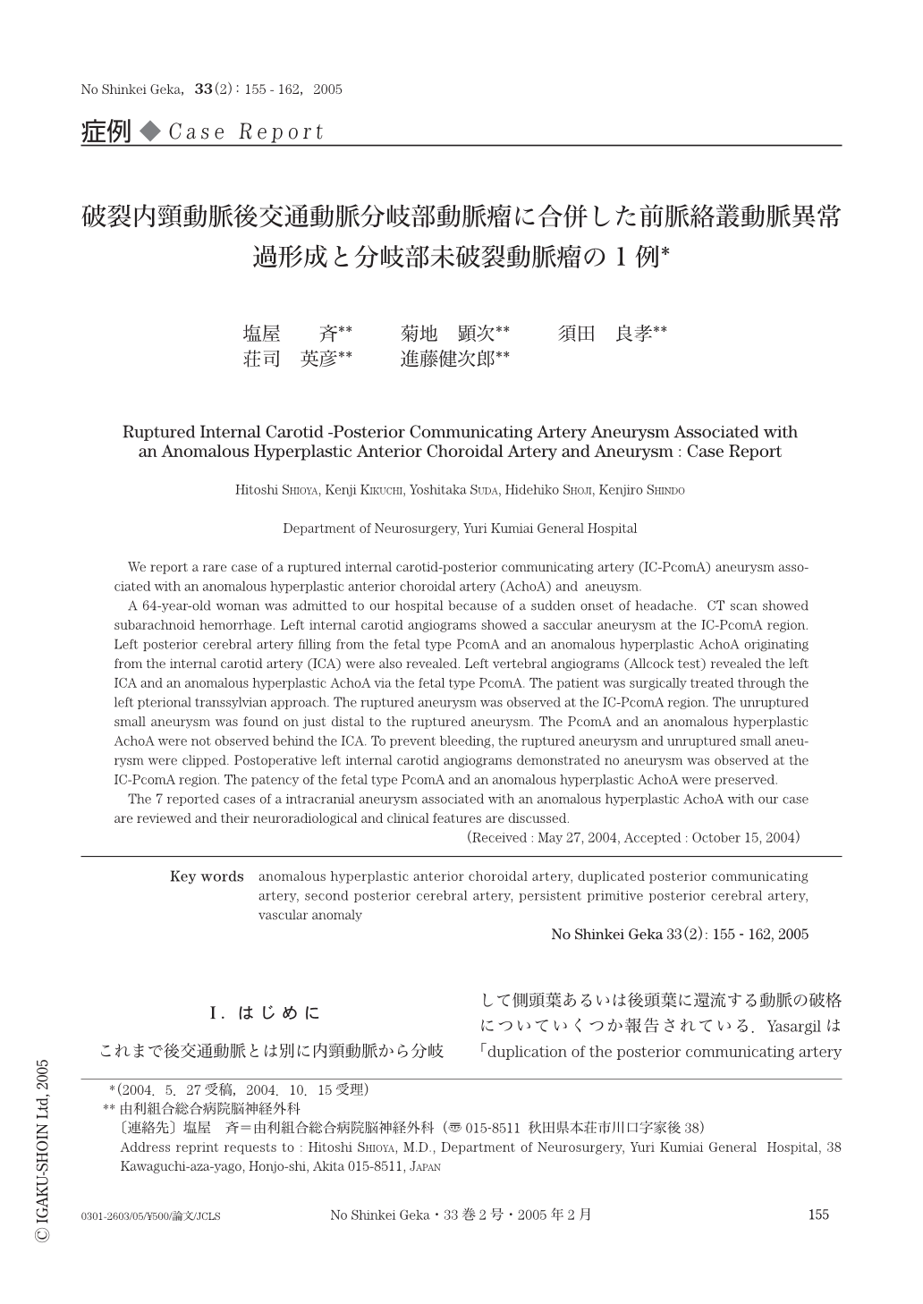
今回,破裂内頸動脈後交通動脈分岐部動脈瘤に合併した前脈絡叢動脈異常過形成と分岐部未破裂動脈瘤の1例を経験したので文献的考察を加えて報告する.
We report a rare case of a ruptured internal carotid-posterior communicating artery (IC-PcomA) aneurysm associated with an anomalous hyperplastic anterior choroidal artery (AchoA) and aneuysm.
A 64-year-old woman was admitted to our hospital because of a sudden onset of headache. CT scan showed subarachnoid hemorrhage. Left internal carotid angiograms showed a saccular aneurysm at the IC-PcomA region. Left posterior cerebral artery filling from the fetal type PcomA and an anomalous hyperplastic AchoA originating from the internal carotid artery (ICA) were also revealed. Left vertebral angiograms (Allcock test) revealed the left ICA and an anomalous hyperplastic AchoA via the fetal type PcomA. The patient was surgically treated through the left pterional transsylvian approach. The ruptured aneurysm was observed at the IC-PcomA region. The unruptured small aneurysm was found on just distal to the ruptured aneurysm. The PcomA and an anomalous hyperplastic AchoA were not observed behind the ICA. To prevent bleeding,the ruptured aneurysm and unruptured small aneurysm were clipped. Postoperative left internal carotid angiograms demonstrated no aneurysm was observed at the IC-PcomA region. The patency of the fetal type PcomA and an anomalous hyperplastic AchoA were preserved.
The 7 reported cases of a intracranial aneurysm associated with an anomalous hyperplastic AchoA with our case are reviewed and their neuroradiological and clinical features are discussed.
Copyright © 2005, Igaku-Shoin Ltd. All rights reserved.