Japanese
English
- 有料閲覧
- Abstract 文献概要
- 1ページ目 Look Inside
I.はじめに
脊髄脂肪腫を構成する脂肪細胞は非常に分化した細胞であり,病理組織学的には,正常脂肪細胞と明かな差異がない2).しかし急速に増大したり2),成人期に発症したりする症例5,8,9,12,14,18,19)が報告されている.一般にcervicothoracic lipoma,lumbosacral lipomaの2群に分けられ,前者は先天奇形との関連は薄く,その意味で腫瘍的性格が強いといわれる.後者は二分脊椎などの先天奇形との関連があり,小児,若年成人に発症するため先天的な腫瘍として知られ12),本来皮下脂肪と連続していることが多く,硬膜内のみに存在するのは稀である4).われわれは成人期発症の腰部硬膜内脂肪腫の稀な1例を経験したので報告する.
A 42-year-old female with intradural lipoma at the level of L1 is reported. She was admitted with a history of 32 months of lumbago, 25 months of pain of the left leg, and 12 months of motor weakness in the left leg. Neurologically, sensory impairment below the L1 der-matome of the left leg, and motor weakness of bilateral legs were noted on admission.
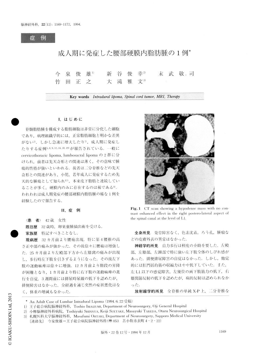
CT demonstrated a low density mass with no con-trast enhanced effect at the level of L1. MRI showed a mass with high signal intensity on the T1-weighted im-age, and low signal intensity on the T2.
L1 laminectomy, and additional Th12 and L2 partial laminectomy were performed and the tumor was par-tially removed. The tumor was completely in the in-tradural space. Pathologically, the tumor consisted of mature adipose cells with normal vessels. Postoper-atively, the epidural effusion at the operative area caused sensory impairment and motor weakness of the right leg.
Finally, the patient came to be neurologically free of defects except for slight sensory diminution of the L4 dermatome of the left leg. In this case, total removal of the tumor was difficult because of adhesion between the tumor, the cauda equina and the conus medullaris. Postoperatively, neurological findings showed a marked improvement. The preoperative neurological deterioration in this case seemed to be associated with simple compression ex-erted on the nerves.

Copyright © 1994, Igaku-Shoin Ltd. All rights reserved.


