Japanese
English
- 有料閲覧
- Abstract 文献概要
- 1ページ目 Look Inside
I.はじめに
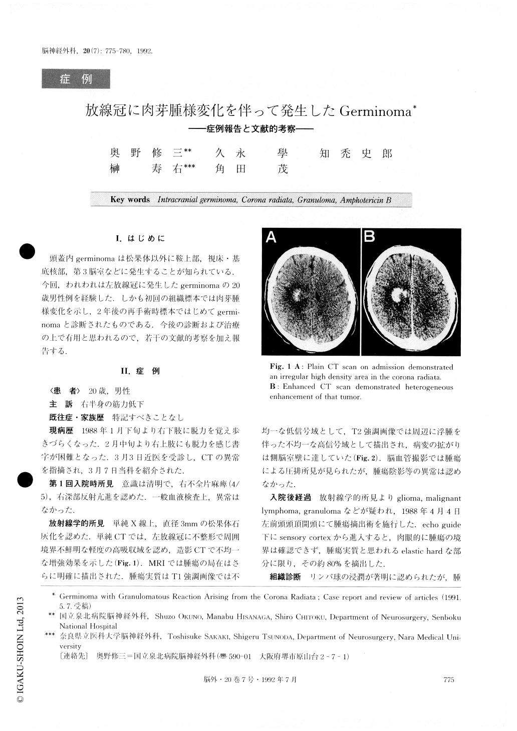
頭蓋内germinomaは松果体以外に鞍上部,視床・基底核部,第3脳室などに発生することが知られている.今回,われわれは左放線冠に発生したgerminomaの20歳男性例を経験した.しかも初回の組織標本では肉芽腫様変化を示し,2年後の再手術時標本ではじめてgermi—nomaと診断されたものである.今後の診断および治療の上で有用と思われるので,若干の文献的考察を加え報告する.
We report a rare case of germinoma with granuloma-tous reaction arising from the corona radiata.
This 20-year-old man was admitted to our hospital complaining of progressive motor weakness on the right side. CT demonstrated a poorly demarcated high density area in the left corona radiata, which was heter-ogeneously enhanced after administration of contrast medium.
Moreover, the continuity of the mass to the ventricu-lar wall was confirmed on MRI. At the first operation, subtotal removal of the tumor was performed through a fronto-parietal craniotomy. .The diagnosis for the speci-fic neoplasm was not established histologically, but gra-nuloma caused by fungal infection was the most likely cause of the lesion. We tried amphotericin B (AmB) and remission of the tumor was obtained. However, during the following 3 months, the size of the tumor gradually enlarged again. AmB was repeatedly adminis-tered, but this time the treatment was ineffective. Six months later, on May 21,1990, the second operation was performed and histological examination revealed typical germinoma consisting of two-cell pattern. Subse-quently, the patient underwent focal irradiation of 33 Gy to the tumor site, and the tumor completely dis-appeared.
As intracranial germinomas are observed to be suc-cessfully cured by radiotherapy and/or chemotherapy, choice of the therapeutic management must be careful-ly determined according to the histological diagnosis, especially in young people. A variety of locations of germinomas and the accompanying granulomatous reactions could create some diagnostic confusion, so great care must he taken in the treatment of much in-tracranial germinomas

Copyright © 1992, Igaku-Shoin Ltd. All rights reserved.


