Japanese
English
- 有料閲覧
- Abstract 文献概要
- 1ページ目 Look Inside
I.はじめに
中耳内に腫瘍が原発することはまれである.その中でも,adenomatous tumorの分類は混乱しており,われわれ脳神経外科医にはなじみが薄い.これまでの報告のほとんどは耳鼻科領域からのものであるが,脳神経外科医が頭蓋底を扱う機会が多くなった現在,錐体骨の病変を扱う際には常に念頭に置くべき疾患である.今回われわれは,中耳内に原発し,組織学的にpapillary patternを呈するadenomatous tumorと診断した症例を経験したので報告する.
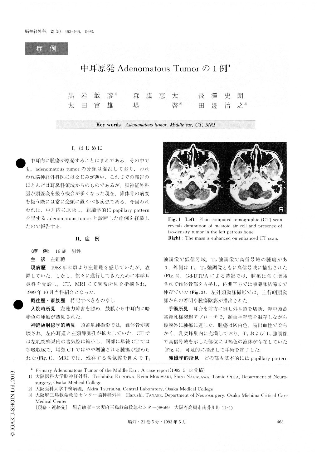
A case of primary adenomatous tumor of the middle ear is described. A 16-year-old male was admitted with a one-year history of left hearing disturbance. Skull X-ray disclosed destruction of the left petrous bone. Computed tomographic scans revealed an iso-dense mass in the left petrous hone, which was slightly enhanced. The tumor appeared as a low intensity mass on T1-weighted magne-tic resonance image (MRI) and as high-intensity mass on T2-weighted MRI, and was enhanced by Gd-DTPA.Fluid in the peritumoral area showed high intensity on Ti-and T2-weighted MRI. External carotid angiography revealed a marked tumor stain fed by the ascending pharyngeal artery. At operation, the tumor was found to be soft and to bleed easily. Histological diagnosis was adenomatous tumor presenting a papillary pattern. Ade-nomatous tumor of the middle ear is rare and difficult to classify. Nevertheless, papillary adenomatous tumor of the middle ear could often be aggressive and malignant in behavior and this patient will require long-term follow up. This is the first report to our knowledge of MRI find-ings about primary adenomatous tumor of the middle ear.
Copyright © 1993, Igaku-Shoin Ltd. All rights reserved.