Japanese
English
- 有料閲覧
- Abstract 文献概要
- 1ページ目 Look Inside
I.はじめに
頭蓋内肉芽腫の原因として,リウマチ性疾患に関連して生じた肉芽腫の報告例は非常に少なく11,13,14),また,そのMRI像に関する報告は認められない.今回,われわれはリウマチ様症状で発症し,11年の経過にわたり寛解と増悪を繰返した頭蓋内多発性の非特異性肉芽腫の1例を経験した.そのMRI所見を中心として,文献的考察も加えて報告する.
We encountered a rare case of a 48-year-old man with intracranial multiple granulomas secondarily caused by rheumatic disease. This was proven surgical-ly after an 11 - year course of remissions and deterio-rations. In 1980, at the age of 32 years, the patient was first seen at the clinic of Neurology of the University Hospital, complaining of swelling and arthralgia of the joints of the knee, ankle, and wrist and with remittent fever and visual disturbance. The patient was dia-gnosed as having possible rheumatoid arthritis, and treated with administration of 30mg/day of predniso-lone, which greatly improved the symptoms. The admi-nistration of 5 to 10mg/clay of prednisolone had been continued after discharge from hospital. In 1985, visual acuity of the left eye decreased, and left facial hypes-thesia developed.
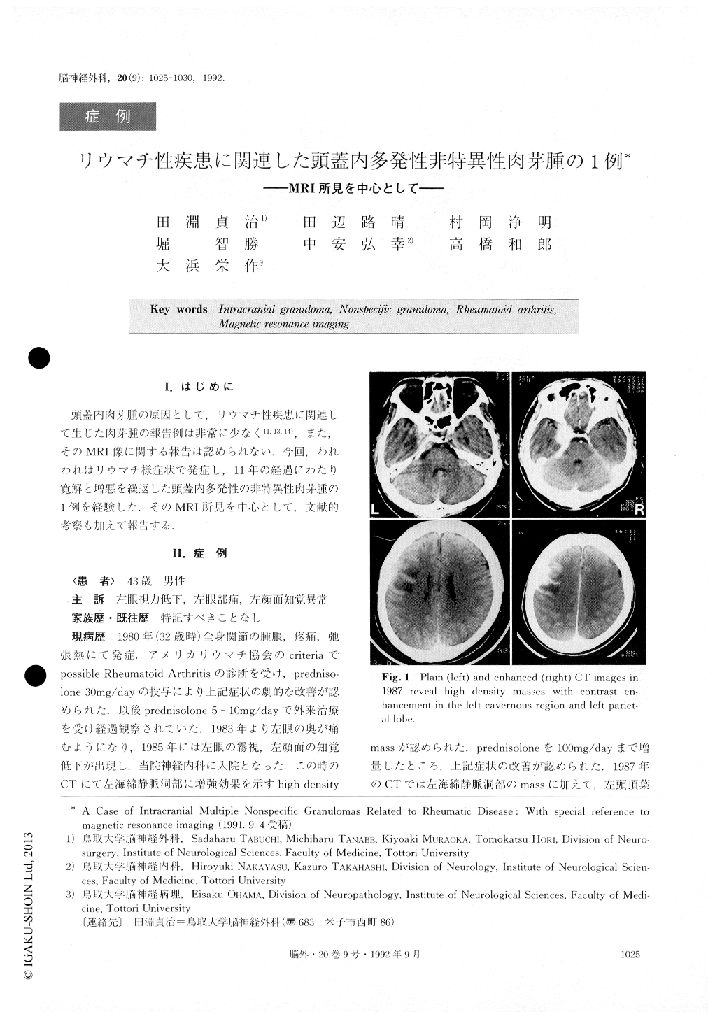
The patient was rehospitalized at the same clinic, and treated with 100mg/day of preclnisolone, which again diminished the symptoms. Computed tomography(CT)on admission showed a high density mass with contrast enhancement in the left cavernous region. In addition to the left cavernous mass, a high density mass was de-tected by CT in the left parietal lobe, in 1987. Visual acuity of the left eye deteriorated in 1989. Because his response to prednisolone had decreased, the visual symptom was treated with gold sodium, which acted effectivity. Symptoms deteriorated again in 1990. Early in 1991, CT and magnetic resonance imaging showed a new mass at the right frontal lobe, while the mass in the left cavernous region had increased in size. The pa-tient was transferred to the clinic of Neurosurgery for surgical treatment.
On admission, left internal carotid angiography showed stenosis at the level of the second cervical vertebra. CT scan showed high density masses in the left cavernousregion, left parietal and right frontal lobes. The areas corresponded with areas of low intensity signals on both T1 - and T2- weighted magnetic resonance im-ages, which were heterogeneously enhanced with gado-linium cliethylenetriamidepentaacetic acid. The mass in the right frontal lobe was subtotally removed, and the mass in the left cavernous region was extirpated includ-ing the dura mater. The removed tissues were histologi-cally of chronic nonspecific granulomatous inflamma-tion, composed mainly of lymphocytes and partially of plasma cells. Considering the clinical course and laboratory and histological findings, the patient was dia-gnosed as having granuloma related to rheumatic dis-ease. Magnetic resonance images of intracranial granu-lomas in the literature were also discussed.
Copyright © 1992, Igaku-Shoin Ltd. All rights reserved.