Japanese
English
- 有料閲覧
- Abstract 文献概要
- 1ページ目 Look Inside
I.はじめに
後大脳動脈瘤は,椎骨脳底動脈領域の動脈瘤の15%14),また全脳動脈瘤の1%14,21),2.7%9)など比較的稀といわれる.今回われわれは,後大脳動脈の,後側頭動脈と頭頂後頭動脈の分岐部,即ちP3部の動脈瘤を経験したので,若干の文献的考察を加えて,これを報告する.
Abstract
A case was reported of surgically treated saccular aneurysm located at the right posterior temporal-parietooccipital artery junction (P3 portion of PCA) . An aneurysm of this portion is said to he rare, and only 7 cases have been described so far.
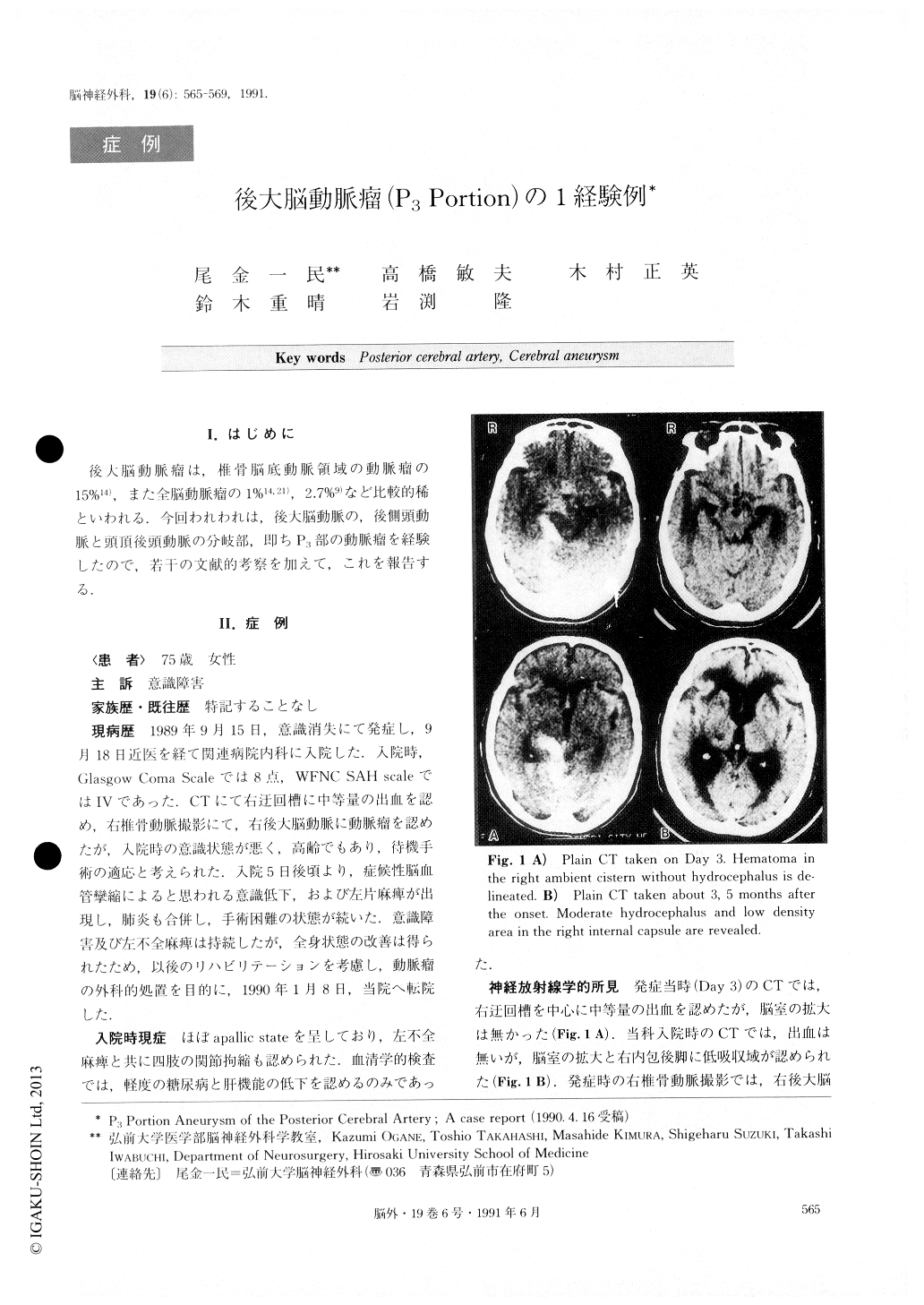
A 74-year-old female was transferred to our clinic, af-ter 3, 5 months of sustaining aneurysmal rupture, for surgical treatment. The patient had been treated conser-vatively because of her severe condition in the early stage. She was in nearly apallic state with left hemi-paresis at the time of admission to our clinic. During the acute stage of her illness, moderate hema-toma in the right ambient cistern without hydrocepha-lus, and an aneurysm at P3 portion of the right post-erior cerebral artery with marked arteriosclerosis were delineated by CT, and by right vertebral angiography respectively. However, in the CT taken 3, 5 months af-ter the onset, moderate hydrocephalus and a low densi-ty area in the right internal capsule were detected.
Aneurysmal neck clipping was performed using the right posterior subtemporal approach, without any de-formity of the parent arteries. Occlusion of the right parietooccipital artery occurred, however, probably on the 2nd postoperative day. Despite the newly developed left homonymous hemianopsia, general condition, in-cluding consciousness level, improved postoperatively particulary after the ventriculo-peritoneal shunt was carried out.

Copyright © 1991, Igaku-Shoin Ltd. All rights reserved.


