Japanese
English
- 有料閲覧
- Abstract 文献概要
- 1ページ目 Look Inside
I.はじめに
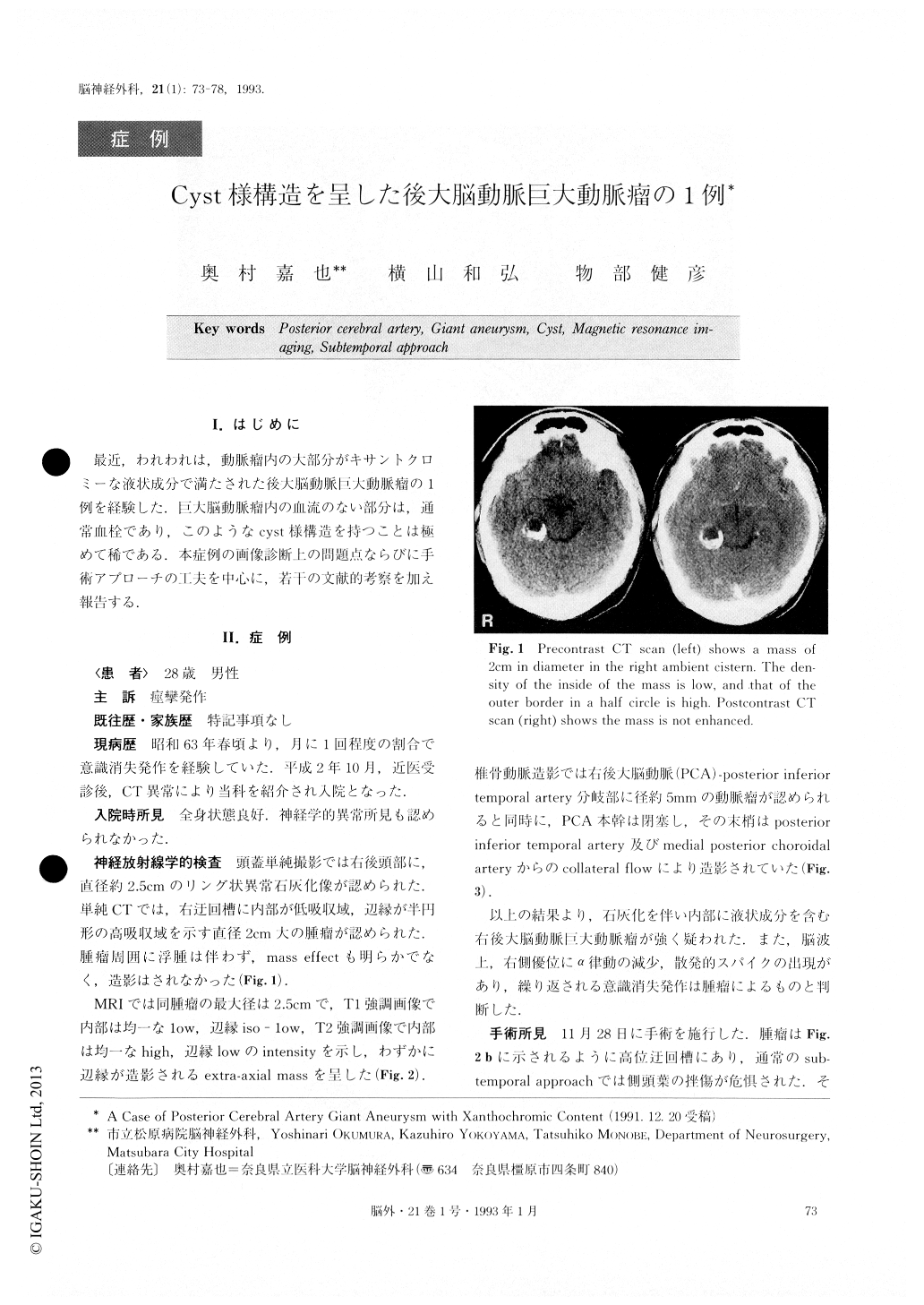
最近,われわれは,動脈瘤内の大部分がキサントクロミーな液状成分で満たされた後大脳動脈巨大動脈瘤の1例を経験した.巨大脳動脈瘤内の血流のない部分は,通常血栓であり,このようなcyst様構造を持つことは極めて稀である.本症例の画像診断上の問題点ならびに手術アプローチの工夫を中心に,若干の文献的考察を加え報告する.
We reported a case of posterior cerebral artery giant aneurysm with xanthochromic content. A 28-year-old male patient was admitted to our department suffering from lapses into unconsciousness once a month for the previous two years and a half. CT scan demonstrated a mass 2cm in diameter in the right ambient cistern. The density of the inside of the mass was low, and that of outer border was high. The mass was not enhanced.MRI demonstrated that the intensity of the inside of the mass was low, and the outer part was iso-low in the Tl weighted image, while the inside was high and the outer part low in the T2 weighted image. Cerebralan-giography showed an aneurysm of 5mm in diameter at the bifurcation of the posterior cerebral artery (PCA) and the posterior inferior temporal artery. There was occlusion of the PCA. Accordingly, we diagnosed it as a calcified giant aneurysm with fluid-like content, and carried out surgery using the extended subtemporal approach. Inside the very stiff yellow-white wall of the aneurysm, there was only xanthochromic fluid without thrombus, and the structure of the aneurysm was cys-tic. The aneurysm was resected following neck clip-ping. After the operation, the patient's lapses into un-consciousness were able to be well controlled with anti-convulsants, and he left our department without any neurological deficit about a month afterwards.The his-tological findings of the specimen show the inner wall of the artery.
Cases of giant aneurysms with xanthochromic con-tent have rarely been reported. Only two are recorded, and this is the first case diagnosed using MRI. Due tothis experience, it is considered that MRI is available for diagnosing giant aneurysms with xanthochromic content, as well as ordinary giant aneurysms. The ex-tended subtemporal approach used in this case is also available when a mass at the high position of the ambient cistern is removed.
Copyright © 1993, Igaku-Shoin Ltd. All rights reserved.