Japanese
English
- 有料閲覧
- Abstract 文献概要
- 1ページ目 Look Inside
I.はじめに
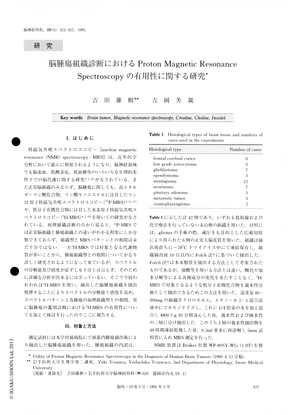
核磁気共鳴スペクトロスコピー[nuclear magneticresonance(NMR)spectroscopy:MRS]は,近年医学分野において盛んに利用されるようになり,脳神経領域でも脳虚血,低酸素症,低血糖等のいろいろな生理的条件下での脳代謝に関する研究2,3,15)がなされている.また正常脳組織のみならず,脳腫瘍に関しても,高エネルギーリン酸化合物,リン酸モノエステルに注目したリン31原子核磁気共鳴スペクトロスコピー(31P-MRS)9,13,17)や,低分子有機化合物に注目した水素原子核磁気共鳴スペクトロスコピー(1H-MRS)4,7,19)を用いての研究がなされている.病理組織診断の点から見ると,31P-MRSでは正常脳組織と腫瘍組織との違いがわかる程度にしか分類できておらず,組織型とMRSパターンとの相関は未だ十分ではない.一方1H-MRSでは対象となる代謝物質が多いことから,腫瘍組織型との相関についてかなり詳しく研究されるようになって来ているが,スペクトルの分解能及び感度が必ずしも十分とは言えず,そのために詳細な分析が出来るには至っていない.そこで今回われわれは1H-MRSを用い,摘出した脳腫瘍組織を抽出処理することによりスペクトルの分解能と感度を高め,スペクトルパターンと各腫瘍の病理組織型との相関,更に脳腫瘍の鑑別診断における1H-MRSの有用性についても加えて検討を行ったのでここに報告する.
Abstract
Proton nuclear magnetic resonance spectroscopy (1H-MRS) was employed in vitro to obtain profiles of va-rious metabolites contained in some of the representa-tive human brain tumors. Extracted samples from each normal and tumoral tissue were used to obtain high-re-solution spectra. Whlie N-acetylaspartate was unequivo-cally demonstrated in normal brain tissues, it could not be detected in brain tumors. Creatine was variably re-duced in brain tumors relative to that in normal brain tissue, and was undetectable in neurinomas and pitui-tary adenomas. Choline was present in all tumors and appeared as relatively prominent peaks in the spectra for meningiomas, pituitary adenomas and metastatic brain tumors. In contrast, levels of creatine and inositol in these tumors were low. Neurinoma showed the largest inositol peak among the tumors examined. Thus compared to normal brain tissue, creatine, choline and inositol in tumors exhibited various degrees of chara-cteristic changes. Therefore we attempted to categorize the spectral patterns of normal and tumoral tissues based on creatine/inositol and/or choline/inositol ratios. There were statistically significant differences between either creatine/inositol ratios and choline/ino-sitol ratios in the tumor groups and the ratios found in normal brain tissue. These ratios also varied relative to different tumor groups.These data suggested that nuc-lear magnetic resonance spectroscopy might yield clini-cally useful information which can not be provided by already widely used magnetic resonance imaging and might be applicable to the differential diagnosis of some brain tumors.

Copyright © 1991, Igaku-Shoin Ltd. All rights reserved.


