Japanese
English
- 有料閲覧
- Abstract 文献概要
- 1ページ目 Look Inside
I.はじめに
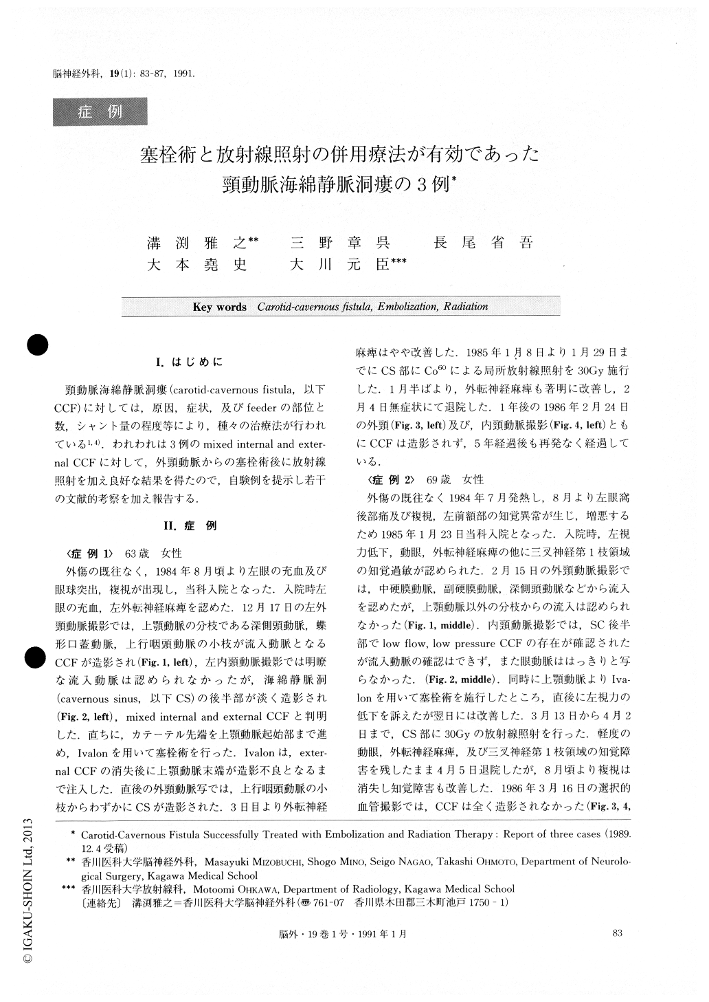
頸動脈海綿静脈洞瘻(carotid-cavernous fistula,以下CCF)に対しては,原因,症状,及びfeederの部位と数,シャント量の程度等により,種々の治療法が行われている1,4).われわれは3例のmixed internal and exter—nal CCFに対して,外頸動脈からの塞栓術後に放射線照射を加え良好な結果を得たので,自験例を提示し若干の文献的考察を加え報告する.
Three cases of mixed internal and external carotid-cavernous fistula (CCF) were successfully treated with embolization of feeders from the external carotid artery (ECA) and focal irradiation to the cavernous sinus (CS).
Cases 1 and 2 were females, 63 and 69 years old re-spectively, both with spontaneous left CCF. Case 3 was a 55 year old male with posttraumatic left CCF.
Symptoms of case 1 were double vision, left chemo- F sis and exophthalmos ; those of case 2 were double vi- sion, left retroorbital pain, left forehead dysesthesia and blurred vision ; and case 3 complained of double vision, left chemosis, left exophthalmos and pulsatile tinnitus. In all three cases, angiography disclosed left CCF fed by ipsilateral dural branches from the internal maxillary artery (IMA) and the internal carotid artery (ICA) . Incase 1, small branches from the ascending pharyngeal artery also fed the CS.
In cases 2 and 3, feeders from the ECA were arising only from branches of the IMA. In case 3, hypertrophy of the meningohypophyseal trunk was visible. In cases 1 and 2, although the CS was opacified, feeders from the ICA were not clearly visible.
Embolizations of branches of the IMA were per-formed in all cases using Ivalon under selective catheterization. In case 1, symptoms partially improved, but in cases 2 and 3, visual symptoms were transiently aggravated.
Focal irradiation to the CS was done with total doses of 30, 30 and 40 Gy each for cases 1, 2, and 3 respec-tively. In case 1, clinical symptoms gradually improved about one third way through irradiation. In case 2, the symptoms improved 4 months after irradiation. Howev-er, in case 3, visual symptoms were rather aggravated by the time 30 Gy irradiation was over. Repeat angio-gram showed prominent hypertrophy of the meningo-hypophyseal trunk and engorgement of the superior ophthalmic vein without recanalization of feeders from the ECA. Additional 10 Gy irradiation was performed. Patient's symptoms gradually improved five months later.
Follow up angiography showed no CCF, 13, 12, and 7 months after irradiation in cases 1, 2, and 3 respec-tively, and no clinical recurrence was recognized over a period of 5, 4, 2.5 years respectively.
It has already been confirmed that even a low dose irradiation with 30 or 40 Gy occludes the CCF com-pletely. Its dose, however, must be the lowest in order to minimize the probability of side effects. Feeders from the ECA should be obliterated first with embo-lization so that following irradiation therapy can be per-formed only for feeders or fistulas from the ICA for the treatment of mixed internal and external CCF. The combined embolization and radiation therapy is indicated in patients with old age, poor risk, surgical in-accessibility, progressive symptoms, and with mixed in-ternal and external low-flow, low-pressure CCF consist-ing of many feeders.
We believe that the combined embolization and irra-diation therapy is probably the most effective alterna-tive with ICA preservation for the mixed internal and external CCF.

Copyright © 1991, Igaku-Shoin Ltd. All rights reserved.


