Japanese
English
- 有料閲覧
- Abstract 文献概要
- 1ページ目 Look Inside
I.はじめに
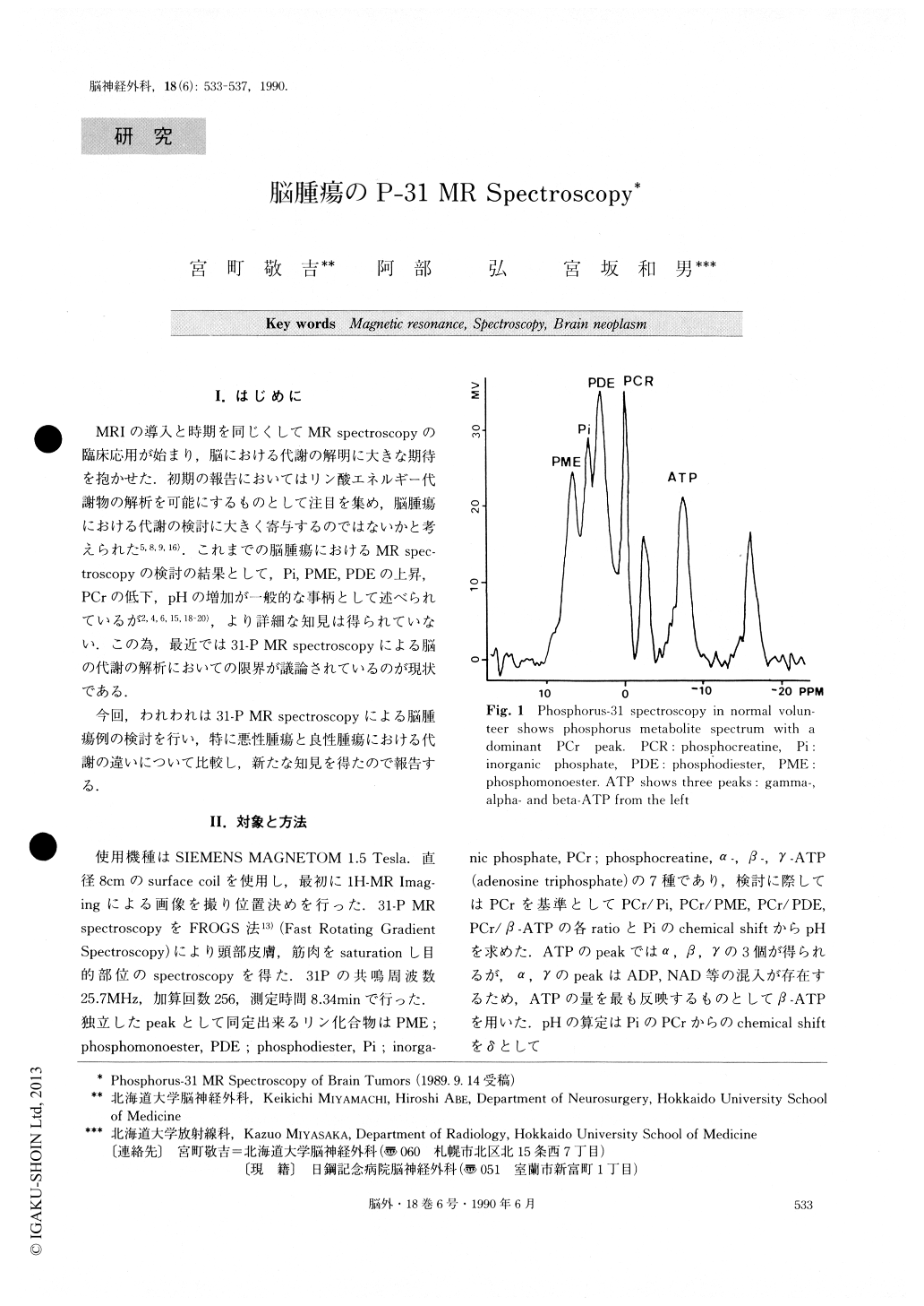
MRIの導入と時期を同じくしてMR spectroscopyの臨床応用が始まり,脳における代謝の解明に大きな期待を抱かせた.初期の報告においてはリン酸エネルギー代謝物の解析を可能にするものとして注目を集め,脳腫瘍における代謝の検討に大きく寄与するのではないかと考えられた5,8,9,16),これまでの脳腫瘍におけるMR spec—troscopyの検討の結果として,Pi,PME,PDEの上昇,PCrの低下,pHの増加が一般的な事柄として述べられているが2,4,6,15,18-20),より詳細な知見は得られていない.この為,最近では31P-MR spectroscopyによる脳の代謝の解析においての限界が議論されているのが現状である.
今回,われわれは31—PMR spectroscopyによる脳腫瘍例の検討を行い,特に悪性腫瘍と良性腫瘍における代謝の違いについて比較し,新たな知見を得たので報告する.
Eleven normal volunteers and 17 subjects with brain tumors were studied by phosphorus-31 Magnetic Reso-nance (MR) spectroscopy. Measurements were per-formed by FROGS (Fast Rotating Gradient Spectroscopy) saturating nearly skin or muscle by Volume of Interest (V0I). This measurement took a-bout 30 minutes for each case, including the imaging procedure, shimming and the measuring of spectro-scopy. In phosphorus-31 MR spectroscopy, pH of the subjects with brain tumors showed a statistically signi-ficant elevation compared with that of the volunteers. In the subjects with benign tumors, only the elevation of pH was significant compared with that in volunteers. There was no other difference. This result suggests that benign tumors have an almost normal metabolic mechanism. Malignant brain tumors showed a decrease of PCr and an increase of P1 and PME. The increase of Pi indicates the increase of energy consumption. The increase of PME is connected with the acceleration of cell proliferation. These findings suggest that, in energy metabolism, there is a difference between malignant tumors arid benign tumors. Phosphorus-31 spectroscopy has the potential to investigate the metabolism in vivo.

Copyright © 1990, Igaku-Shoin Ltd. All rights reserved.


