Japanese
English
症例
6年間の経過観察でCystの増大がCT上確認された脳有鉤嚢虫症の1例
A Case of Cerebral Cysticercosis: Cyst growth is confirmed by CT scan during 6 years of follow-up
寺田 耕作
1
,
妹尾 包人
1,3
,
上津原 甲一
1
,
朝倉 哲彦
2
Kousaku TERADA
1
,
Kanehito SENO
1,3
,
Koichi UETSUHARA
1
,
Tetsuhiko ASAKURA
2
1鹿児島県立大島病院脳神経外科
2鹿児島大学脳神経外科
3大井病院脳神経外科
1Department of Neurosurgery, Oshima Prefectural Hospital of Kagoshima
2Department of Neurosurgery, Kagoshima University
キーワード:
Cerebral cysticercosis
,
CT
,
Cyst growth
Keyword:
Cerebral cysticercosis
,
CT
,
Cyst growth
pp.391-395
発行日 1990年4月10日
Published Date 1990/4/10
DOI https://doi.org/10.11477/mf.1436900064
- 有料閲覧
- Abstract 文献概要
- 1ページ目 Look Inside
I.はじめに
人体有鉤嚢虫症は有鉤条虫(Taenia solium)の幼虫である有鉤嚢虫(Cysticercus cellulosae)が全身の諸臓器に寄生するために惹起される疾患であり,脳神経外科領域においても脳有鉤嚢虫症として古くより知られている16)
今回われわれは,6年の経過にてcystの増大をCT上確認し外科的に摘出しえた脳有鉤嚢虫症の1例を経験したので,若干の文献的考察を加え報告する.
Abstract
A rare case of a 40-year-old woman with cerebral cysticercosis is reported. She has lived on the Island of Tokunoshima and has never travelled overseas.
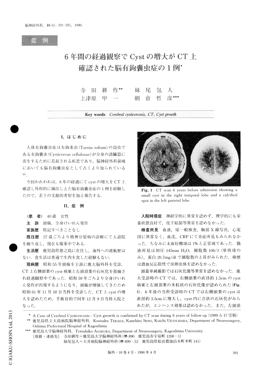
She was hospitalized in the hospital of Kagoshima University in 1980. A small cyst in the right temporal lobe, and a calcified area in the left parietal lobe was noticed on CT scan. When she was admitted to our hospital in December 1986, the cyst in the right temporal lobe was larger than it was 6 years before.

Copyright © 1990, Igaku-Shoin Ltd. All rights reserved.


