Japanese
English
- 有料閲覧
PPV購入案内
1,320円 (1,200円+税10%) こちらは、1223ページから1228ページの文献です。購入には医書.jp本会員登録が必要です。
会員登録完了後に改めて上記「購入サイトへ」を選択してください。
または「購入サイトへ」選択後に「購入ログイン」、「新規会員登録」の順に進んでください。
会員登録について詳しくはをご確認ください。
- Abstract 文献概要
- 1ページ目 Look Inside
Ⅰ.はじめに
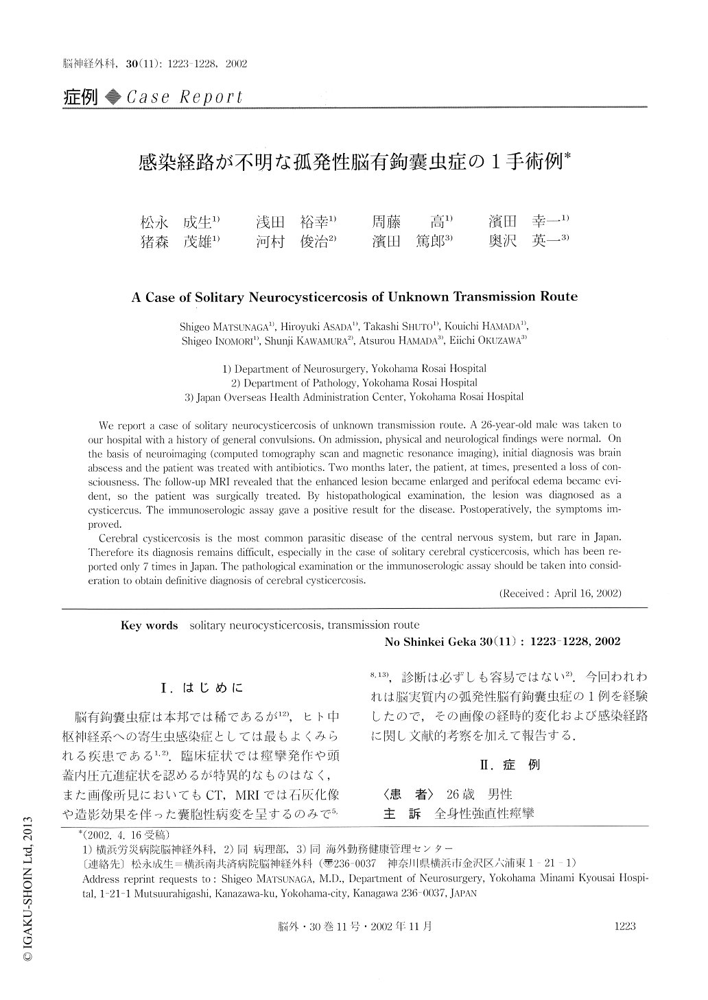
脳有鉤嚢虫症は本邦では稀であるが12),ヒト中枢神経系への寄生虫感染症としては最もよくみられる疾患である1,2).臨床症状では痙攣発作や頭蓋内圧亢進症状を認めるが特異的なものはなく,また画像所見においてもCT,MRIでは石灰化像や造影効果を伴った嚢胞性病変を呈するのみで5,8,13),診断は必ずしも容易ではない2).今回われわれは脳実質内の弧発性脳有鉤嚢虫症の1例を経験したので,その画像の経時的変化および感染経路に関し文献的考察を加えて報告する.
We report a case of solitary neurocysticercosis of unknown transmission route. A 26-year-old male was taken to our hospital with a history of general convulsions. On admission, physical and neurological findings were normal. On the basis of neuroimaging (computed tomography scan and magnetic resonance imaging), initial diagnosis was brain abscess and the patient was treated with antibiotics. Two months later, the patient, at times, presented a loss of con-sciousness. The follow-up MRI revealed that the enhanced lesion became enlarged and perifocal edema became evi-dent, so the patient was surgically treated.
Copyright © 2002, Igaku-Shoin Ltd. All rights reserved.